Canine intraocular neoplasia

37 Canine intraocular neoplasia


CLINICAL EXAMINATION


Since most cases of intraocular neoplasia will be primary rather than secondary it is likely that the general clinical examination will be normal. Obviously if both eyes are affected or the patient is unwell, then metastatic disease is more likely. However, the majority of cases will be unilateral. As such, the abnormal eye can be compared to the normal one during examination which is helpful.


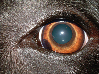
The affected eye might be blind but usually vision is present. Thus a menace response is likely to be normal. Some conjunctival hyperaemia and episcleral congestion might be present but not always. Normally a discoloured lesion will be present. This can be on or in the iris, or behind it poking thorough the pupil. Its colour can vary from dark brown to pale pink and various shades in between (Figure 37.1). The pupil is frequently distorted (dyscoria) but pupillary light reflexes remain. Mild uveitis, with aqueous flare and episcleral congestion, can be present and occasionally the patient is not presented until the eye is painful and enlarged through secondary glaucoma.


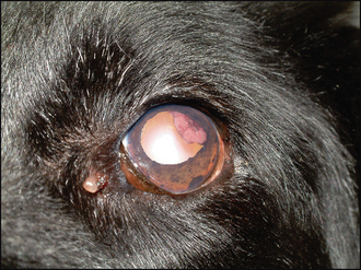
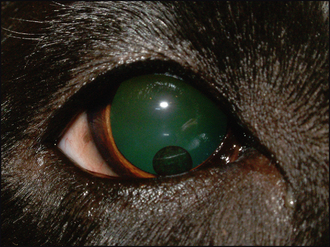
If the neoplasm is based in the iris, then the discolouration will be visible within the iris, and can be raised above it (Figure 37.2). Thus looking at the eye from the side can be useful to determine if the lesion protrudes out from the plane of the iris. Ciliary body-based masses are more difficult to see initially; however, by dilating the pupil the lesion should be more easily visualized through the pupil, especially when looking tangentially across the eye. Vitreal haze is often present with ciliary body neoplasms. Fundus neoplasia is rare but occasionally chor-oidal extension of uveal melanomas does occur.


The most commonly encountered uveal tumour in dogs is the iris melanoma/melanocytoma, with ciliary body adenoma/adenocarcinoma the next most frequent. However, many tumour types can occur – haemangiosarcoma, medulloepithelioma, primary lymphoma and so on, and although the clinical examination can suggest the tumour type, it is only on histological examination or cytology that this can be confirmed.


CASE WORK-UP


If an ocular mass is suspected a thorough clinical examination is required to try to establish whether this is primary or secondary. Thus if there are no changes in the fellow eye, nor on general examination, it is appropriate to assume the lesion is primary – especially if it presents as a mass rather than a diffuse uveitis.


Gonioscopy can be useful to determine whether extension into the drainage angle has occurred (which could indicate an increased risk of metastasis as tumour cells escape from the eye into the general circulation). Ocular ultrasonography can be considered but this might not provide further information about the lesion (although it can differentiate a solid mass from a cyst). Chest and abdominal radiography and/or ultrasonography should be undertaken if there is any suspicion of metastatic spread – for example, in advanced cases where there is not only a visible mass but also associated uveitis and secondary glaucoma.


Fine needle aspirates of draining lymph nodes can also be performed should metastatic disease be suspected. However, since many cases of ocular neoplasia in dogs are primary, and metastasis is uncommon, extensive work-up is not usually required unless there is an unusual aspect to the presentation.


Stay updated, free articles. Join our Telegram channel

Sep 10, 2016 | Posted by in SMALL ANIMAL | Comments Off on Canine intraocular neoplasia

Full access? Get Clinical Tree

Get Clinical Tree app for offline access