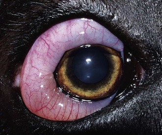
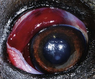
Canine Conjunctivitis

Chapter 244


Canine Conjunctivitis


Conjunctivitis is a common disorder in dogs. The causes of canine conjunctivitis are numerous, including primary conjunctival diseases and other extraocular, intraocular, and systemic conditions that lead to secondary conjunctivitis. Clinical manifestations of conjunctivitis frequently are nonspecific and may be similar in spite of diverse causes. Furthermore, severe ocular diseases and potentially life-threatening systemic conditions can present initially as conjunctivitis. Failure to consider both local and systemic causes of conjunctivitis can have dire consequences for ocular and systemic health; therefore a methodical clinical approach to conjunctivitis should be followed.


Clinical Signs


Conjunctivitis invariably is associated with some combination of the following clinical signs: ocular discharge, chemosis, hyperemia, discomfort, pruritus, tissue proliferation, ulceration, and hemorrhage. Ocular discharge may be characterized by serous, mucoid, mucopurulent, or serosanguineous fluids. Epiphora, overflow of serous tears, results from excessive production or inadequate nasolacrimal drainage. Chemosis appears as conjunctival swelling and is the clinical manifestation of conjunctival edema caused by increased vascular permeability and fluid extravasation (Figure 244-1). Hyperemia is a red conjunctival discoloration (“red eye”) and is a clinically observable manifestation of vasodilation and increased blood flow. Ocular discomfort and pruritus most commonly manifest as blepharospasm and periocular rubbing.


Tissue proliferation is divided into two distinct types: lymphatic and epithelial. Lymphatic proliferation frequently is termed follicular conjunctivitis and appears as small, round, semitransparent, elevated lesions representing lymphocytic aggregates. The occasional occurrence of conjunctival lymphatic follicles on the posterior aspect of the nictitans membrane is normal; however, increased numbers of follicles or their presence in other anatomic locations is pathologic. Epithelial hyperplasia or keratinization results in irregular, opaque, pink to red, elevated lesions of variable size. Both lymphatic and epithelial conjunctival tissue proliferation are indicative of chronic inflammation but are otherwise nonspecific clinical findings.


Ulceration of the conjunctival epithelium may occur with any severe conjunctivitis but is most common in conjunctivitis of viral or traumatic origin. Conjunctival ulcers appear as flat, irregular, pale or pink regions on the conjunctival surface that retain fluorescein stain and are surrounded by a hyperemic border. Hemorrhage may occur in the conjunctival epithelium or subconjunctival space. Both intraconjunctival and subconjunctival hemorrhages appear as bright or dark red regions of variable shape and size (Figure 244-2). Conjunctival hemorrhage is frequently detected in dogs with traumatic or viral conjunctivitis but also can be a manifestation of systemic disease, including coagulopathy, hypertension, hyperviscosity, platelet disorders, and vasculitis.


Clinical Evaluation


The clinical approach to canine conjunctivitis is similar to the approach to any nonspecific ocular lesion that can have various causes, including investigation of both primary and secondary causes of conjunctival inflammation. A thorough history taking, physical examination, and ocular examination are performed to identify the specific cause when possible or to narrow the differential diagnosis and exclude more serious causes of conjunctivitis such as systemic or intraocular disease. Historical details to collect include the onset and duration of clinical signs, presence and nature of ocular discharge, changes in vision, presence of pain or pruritus, and known concurrent systemic and ocular diseases. All ocular and systemic drug treatments should be identified, including over-the-counter medications and medications administered by the client on his or her own accord. It is beneficial to request that clients not clean ocular discharge from the dog before the evaluation. A complete physical examination is performed on all dogs with conjunctivitis, including evaluation of body temperature, thoracic auscultation, oral cavity examination, and regional lymph node palpation.


A complete ophthalmic examination always is indicated for dogs with conjunctivitis, including Schirmer tear tests, ocular surface fluorescein staining, and tonometry (see Chapter 242). This simple examination strategy maximizes the chances of correctly identifying the cause of the conjunctivitis and prevents missing more serious ocular diseases that initially may resemble primary conjunctivitis. These include ulcerative keratitis, uveitis, and glaucoma. Examination should begin with a general evaluation of facial conformation and the size, position, and symmetry of the globe, orbits, eyelids, and pupils. The general evaluation is most productive when performed at a distance from the dog’s head and with minimal restraint. Palpation of the periocular facial regions and adnexa is performed and menace responses, palpebral reflexes, pupillary light reflexes, ocular motility, and globe retropulsion are assessed.


Detailed examination of the adnexa, ocular surface, and intraocular structures is then performed with magnification and a bright focal light source. Eyelid conformation is assessed and the eyelid margins and conjunctiva are methodically examined to detect foreign bodies, cilia abnormalities, and other lesions. The bulbar surface of the nictitans membrane is examined for foreign material or the presence of abundant lymphoid follicles. Examination of the anterior segment of the globe may be performed without dilating the pupil; however, complete evaluation of the lens and posterior segment can be accomplished only with pharmacologic mydriasis.


Causes and Management of Canine Conjunctivitis


As indicated earlier, the causes of conjunctivitis include primary conjunctival diseases, secondary manifestations of other ocular diseases, and secondary manifestations of systemic diseases. Primary causes of conjunctivitis are those in which the disease process is limited to the conjunctiva and include allergic, frictional irritant, immune-mediated, infectious, and traumatic conditions.


Primary Conjunctival Diseases


Allergic Conjunctivitis


Conjunctivitis associated with allergic conditions occurs frequently in the dog and can be divided into three general types: atopic conjunctivitis, drug reaction conjunctivitis, and conjunctivitis caused by insect envenomation. Atopic conjunctivitis often is accompanied by atopic dermatitis but can occur alone. Dogs frequently show mild and seasonal hyperemia, chemosis, epiphora, and ocular pruritus. Conjunctival follicle formation occurs in chronic cases. Atopic conjunctivitis generally is a diagnosis of exclusion; however, atopic dermatitis and seasonality are suggestive of this cause. Allergen immunotherapy and allergen avoidance are definitive treatments for atopic conjunctivitis but may be impractical for some dogs. The use of topical ophthalmic corticosteroids applied two or three times daily for short durations (1 to 2 weeks) as needed for flare-ups generally is effective in controlling clinical signs. Dogs with recurrent episodes, a protracted clinical course, or contraindications to topical corticosteroids benefit from long-term or lifelong topical ocular cyclosporine therapy (0.2% to 2% solution or ointment, applied twice daily). Continual cyclosporine therapy generally reduces or eliminates the need for pulse therapy with topical corticosteroids.


Drug reaction conjunctivitis is a hypersensitivity reaction that often results in severe clinical signs. Concurrent blepharitis, often with dermal ulceration, frequently is present. Conjunctivitis may develop at any time during drug use and with any medication. Ophthalmic medications containing neomycin and carbonic anhydrase inhibitors are among the most common causes of allergic conjunctivitis in dogs. Treatment consists of discontinuing all ophthalmic medications for 1 to 2 weeks (when possible) and slowly reintroducing medications individually until the offending pharmaceutical is identified. Topical corticosteroid therapy can assist in relieving clinical signs, but a medication that is unlikely to be associated with this type of reaction should be chosen (e.g., prednisolone acetate 1% solution).


Insect bites and stings anywhere on the body—not just on periocular tissues—may result in dramatic conjunctivitis. This form of conjunctivitis typically has a rapid onset and is characterized by severe bilateral chemosis with the conspicuous absence of other acute clinical signs of conjunctivitis (see Figure 244-1). Therapy consists of a single dose of systemic corticosteroid and possibly an antihistamine followed by several days of topical corticosteroid administration. Rapid resolution of the chemosis is typical.

< div class='tao-gold-member'>

Only gold members can continue reading. Log In or Register to continue

Stay updated, free articles. Join our Telegram channel

Jul 18, 2016 | Posted by in PHARMACOLOGY, TOXICOLOGY & THERAPEUTICS | Comments Off on Canine Conjunctivitis

Full access? Get Clinical Tree

Get Clinical Tree app for offline access