Brachycephalic pigmentary keratitis

28 Brachycephalic pigmentary keratitis


CLINICAL EXAMINATION


General clinical examination is usually normal. However, multiple, often quite subtle, abnormalities are detected on ophthalmic examination. The most striking feature is a film of dark pigment extending over the corneas. This is usually bilateral, but not necessarily symmetrical. It is normally most dense ventromedially and extends in a triangle with the apex centrally (Figure 28.1).
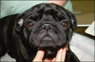


However, other less obvious changes are likely to be present. The dog can probably see, so menace responses will be present, but these might be reduced, especially if directing the threatening gesture from a ventral or ventromedial direction. Palpebral reflexes should be checked – they are likely to be normal in that the animal will blink, but the eyelids might not fully meet on blinking due to some degree of lagophthalmos. Blink rates might be reduced and incomplete or partial blinks are not uncommon. It is sensible to ask the owners whether the dog sleeps with his eyes fully closed – sometimes they do not. Schirmer tear test readings should be evaluated. These could be normal, increased (especially if corneal ulceration is present) or reduced. Low grade keratoconjunctivitis sicca is not infrequent in brachycephalic dogs.


Careful evaluation of the lid margins should be undertaken. A ventromedial entropion is often present. The eyelid margins will appear tight against the cornea, with hairs from the lid touching the corneal surface. The third eyelid is not normally elevated or providing any corneal protection. Hairs are often also present on the caruncles – the small fleshy protuberances at the medial canthi – and these can both irritate the cornea and act as a wick to draw tears down the face rather than down the nasolacrimal punctae.


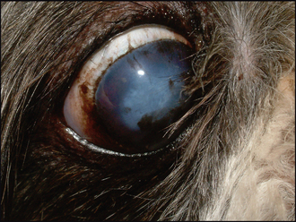
Nasal fold trichiasis can be present. Here the hairs from the skin over the nasal folds are in contact with the corneal surface. Viewing the patient from the side and above as well as directly in front will help to determine whether significant trichiasis is present. Close evaluation of the nasolacrimal punctae should be performed. These are normally present, and of normal size, but the medial entropion causes the openings to be physically closed (like a straw flattening on itself). This will contribute to any epiphora. The skin ventromedial to the eye should be checked – the epiphora could allow a low grade moist dermatitis to develop here. Distichia can be present – commonly in English bulldogs for example – but as these are often subclinical they must be carefully examined to see if they are actually in contact with the cornea or the tear film and are truly contributing to the corneal disease, or whether they are purely an incidental finding (Figure 28.2).


Conjunctival hyperaemia is not a consistent finding – it will be present if there is concurrent corneal ulceration but otherwise the conjunctiva is often normal. Detailed corneal examination is required. The pigment is normally superficial but can be quite thick in places. It will almost always be associated with some degree of corneal vascularization – this is best appreciated at the leading edge of the pigment since the dark colouration will obscure any blood vessels beneath it. If ulceration has occurred previously it is most common centrally in brachycephalics and so some white stromal scarring and/or irregular corneal outline might be present in this area. Active ulceration can be shallow or quite deep with a marked vascular reaction and all the signs typically present with any ulceration. It goes without saying that fluorescein testing is mandatory. As usual, the excess dye should be carefully flushed away using sterile saline solution – the surface of the cornea is likely to be irregular due to the pigment and previous scarring, and pooling of dye in these patches could lead to false positives.


Although intraocular contents are usually normal, evaluation of them can be difficult. Distant direct ophthalmoscopy can be used to assess pupil size and gain a tapetal reflex (albeit reduced). Indirect ophthalmoscopy offers the best way of evaluating the fundus since the view through semi-opaque ocular media is superior with this method over direct ophthalmoscopy.


CASE WORK-UP


Once a thorough ocular examination has been performed to determine the underlying reason for the pigment deposition, then a management plan can be drawn up. This will probably include both surgical and medical options and if the former is indicated a full general examination should be undertaken. Particular attention should be paid to the presence of other brachycephalic problems – notably upper airway conditions – which might require surgery as well as the eyes, or at least should be borne in mind when planning general anaesthesia. Apart from this, there is limited work-up necessary since the ophthalmic examination should be sufficient to reach a definitive diagnosis.


Stay updated, free articles. Join our Telegram channel

Sep 10, 2016 | Posted by in SMALL ANIMAL | Comments Off on Brachycephalic pigmentary keratitis

Full access? Get Clinical Tree

Get Clinical Tree app for offline access