28 Brachycephalic pigmentary keratitis
CLINICAL EXAMINATION
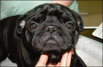
General clinical examination is usually normal. However, multiple, often quite subtle, abnormalities are detected on ophthalmic examination. The most striking feature is a film of dark pigment extending over the corneas. This is usually bilateral, but not necessarily symmetrical. It is normally most dense ventromedially and extends in a triangle with the apex centrally (Figure 28.1).

Figure 28.1 Bilateral pigmentary keratitis in a young Pug. Note the ventromedial pigment in both corneas.
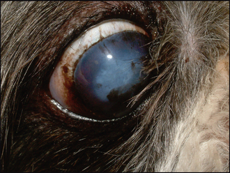
Nasal fold trichiasis can be present. Here the hairs from the skin over the nasal folds are in contact with the corneal surface. Viewing the patient from the side and above as well as directly in front will help to determine whether significant trichiasis is present. Close evaluation of the nasolacrimal punctae should be performed. These are normally present, and of normal size, but the medial entropion causes the openings to be physically closed (like a straw flattening on itself). This will contribute to any epiphora. The skin ventromedial to the eye should be checked – the epiphora could allow a low grade moist dermatitis to develop here. Distichia can be present – commonly in English bulldogs for example – but as these are often subclinical they must be carefully examined to see if they are actually in contact with the cornea or the tear film and are truly contributing to the corneal disease, or whether they are purely an incidental finding (Figure 28.2).
CASE WORK-UP
Once a thorough ocular examination has been performed to determine the underlying reason for the pigment deposition, then a management plan can be drawn up. This will probably include both surgical and medical options and if the former is indicated a full general examination should be undertaken. Particular attention should be paid to the presence of other brachycephalic problems – notably upper airway conditions – which might require surgery as well as the eyes, or at least should be borne in mind when planning general anaesthesia. Apart from this, there is limited work-up necessary since the ophthalmic examination should be sufficient to reach a definitive diagnosis.
Stay updated, free articles. Join our Telegram channel

Full access? Get Clinical Tree


