Basics of Exercise Physiology


Basics of Exercise Physiology


Robert Gillette and R. Barry Dale


image


A good basic knowledge of exercise physiology is necessary when designing and implementing a rehabilitation or conditioning program. The definition of training, for the purposes of this chapter, refers to working with the dog in an obedience training program that addresses behavioral aspects, whereas conditioning refers to an exercise or workout program that addresses the animal’s physiologic capacity to perform work. Exercise physiology is a discipline that examines how exercise affects the body and is applicable in the field of physical rehabilitation.


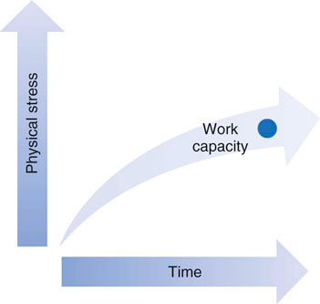
The level of conditioning of a dog constantly changes any time the body performs at a specific metabolic level proportionate to its current adaptation to physical stress (Figure 8-1). The systems of the body are conditioned to maintain homeostasis at this level. If the demands placed on the body increase over time, the body adapts and conditions itself to maintain homeostasis at this new level. An understanding of the physiologic changes involved in this process helps in the development of programs that allow regulation of the conditioning or reconditioning of the body. These programs may be used to condition the entire body or to focus on individual body segments. In physical rehabilitation, programs may be developed to treat injuries, to stimulate healing of the injuries, and to enhance reconditioning of the injured segment. Because of the biomechanical forces acting on the body during the reparative phases of healing, certain segments may become stronger than other segments. Exercise programs should be implemented to remedy any conditioning imbalances of the body. The basic goals should be to allow the tissues to heal, then to recondition the tissues so that they can accept predetermined workloads, and finally to recondition the entire body to balance any conditioning deficits.


Cardiopulmonary Physiology


The circulatory system carries various oxygen, substrates, and chemical messengers; removes carbon dioxide and metabolic byproducts; assists with thermoregulation; and is an important mediator for acid-base balance.1


Blood consists of fluid and cellular components. Blood plasma is the liquid portion of blood and consists of water, proteins, electrolytes, dissolved gases, and nutrients.1 The cellular constituents consist of erythrocytes, leukocytes, and platelets. Oxygen and carbon dioxide (discussed in the next section) dissolve to a limited extent in blood plasma, but are largely carried bound to hemoglobin found in erythrocytes.


The heart serves to pump blood to the pulmonary and peripheral circulation. Resting heart rate is approximately 80 beats per minute for a large dog.1 In response to exercise, heart rate and myocardial contractility increase, which concomitantly increases cardiac output. A large dog typically has increases of 30% for stroke volume, 300% for heart rate, and approximately 400% for cardiac output during vigorous exercise when compared with rest.1


Muscle Physiology


Muscle is a tissue greatly affected by exercise or disuse associated with injury. Understanding muscle function and the molecular events of muscle contraction provides a basis for the concepts of physical rehabilitation and exercise physiology. There are three types of muscle in the body: skeletal, smooth, and cardiac. Skeletal muscles connect one bone to another. Each muscle consists of thousands of myocytes. A muscle fiber is long, fusiform, and surrounded by a plasma membrane called the sarcolemma. Inside these fibers are myofibrils, which consist of filaments composed of contractile proteins. These contractile proteins are arranged in units called sarcomeres.


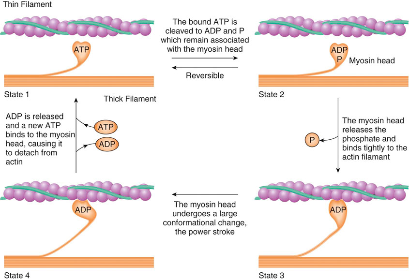
Actin and myosin are two types of protein chains in the sarcomere. They interact as a result of enzymatic and chemical reactions to produce muscle contraction. Calcium and phosphate are chemical components involved with the production of muscle contractions. Phosphate is in the form of adenosine triphosphate (ATP). ATP binds to a receptor site at the end of the myosin leverage arm. The actin filament includes troponin, which is bound to strands of tropomyosin. A calcium ion attaches to the troponin molecule, which changes the shape of the tropomyosin. This action opens a myosin-specific binding site on the actin protein chain. Energy is emitted when ATP releases a phosphate ion, producing adenosine diphosphate (ADP). The resultant energy allows the ADP to create a bond between the open binding site on the actin filament and the myosin leverage arm. This bond changes the myosin structure, allowing leverage to produce a contraction between the two fibers. The ADP is released and the lever arm is freed. Energy is then required to add a phosphate group to the ADP, re-creating ATP, which is then used for further contractions (Figure 8-2). The accumulated contractions of the muscle fibers create contraction of an entire muscle.


Muscle Structure and Function


The innervation to a muscle controls the muscle contraction by directing an action potential along an efferent motor neuron. A single motor nerve diverges to innervate many muscle fibers. The combination of a nerve and the muscle fiber it innervates is called a motor unit. Contraction of an entire muscle is a result of the cumulative contraction of many motor units.


The muscle fibers are grouped together and organized with other fibers by a sheath of connective tissue that is named according to its level of organization. Endomysium covers each of the muscle fibers themselves, and perimysium separates discrete bundles of fibers. Epimysium is the connective tissue layer that surrounds the grouped bundles. These three layers of connective tissue are longitudinally continuous throughout the muscle belly and blend together at each end with the proximal and distal tendons. Muscle fascia is the sheath that covers the epimysium and serves to protect each muscle from movement over hard structures or movement from adjacent muscles. The arrangement of these fibers plays a role in the function of each muscle. It is the combination of motor unit group, the characteristics of the muscle fibers, and fiber arrangement that dictates the resultant type of muscular function (strength, power, or endurance).


In general, muscle contraction and work are transferred through the tendon and its attachment to a bone. The musculotendinous junction is a layered transition between muscle fibers and the collagen of the tendons. Tendons of origin and insertion may run throughout the length of the structure. Musculotendinous structures are closely tied to functional requirements. Structural shapes of muscle include fusiform and pennate forms. Pennate muscles may be divided into unipennate, bipennate, and multipennate forms. Pennate structures allow a muscle to lift great loads but through a small range of motion, such as in vertebrae. Fusiform structures have the ability to lift a small load at a great velocity through a large range of motion. These types of muscles include the biceps brachii and brachialis muscles in the antebrachium. These two forms of muscle shapes can work together if both strength and speed of movement are needed in a particular joint, such as the shoulder or hip joint.


Muscle Energy Systems


Muscles require energy to maintain basal metabolism and additional energy during physical activity. The body uses three systems to provide this energy: (1) immediate energy sources, (2) glycolytic metabolism, and (3) oxidative metabolism. The type of activity the muscle is performing determines which of the systems will be used. The cellular environment must also be conducive to these processes. Factors affecting the cellular environment include pH, hydration status, temperature, and presence of the proper enzymes. Changes in any of these factors may alter the reactions needed for energy production. While energy production is occurring, metabolic byproducts are simultaneously produced. Byproduct removal is essential, because local accumulation of these substances alters the cellular environment.


The immediate energy source involves intracellular ATP, creatine phosphate (CP), and the ADP-myokinase reaction to provide energy for activity. Intracellular ATP is the first energy source used for contraction. A limited amount of ATP is stored at the myosin crossbridges, near mitochondria, and beneath sarcolemma. ATP breakdown is a hydrolytic reaction and is highly regulated. The body does not allow large changes in ATP content. CP is a high-energy compound used to ensure that the ATP concentration does not become depleted. It is located near the actin-myosin filaments and in the mitochondrial membrane. The CP molecule donates a phosphate group to ADP to replenish the ATP used at the contraction site. The third immediate energy source comes from the myokinase reaction. Myokinase is the enzyme that allows two ADP molecules to combine to form ATP and adenosine monophosphate (AMP). This reaction provides little energy to the system, but AMP serves as one of the allosteric modulators to stimulate carbohydrate (glucose) breakdown in glycolytic metabolism. Energy from this system lasts anywhere from 5 to 10 seconds with high-intensity exercise and occasionally up to 20 seconds in some elite athletes.


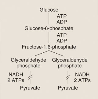
The glycolytic pathway provides energy from 5 to 20 seconds up to 2 minutes as a result of the anaerobic breakdown of glucose. This is a more complex form of energy production, using multiple enzymes and reactions. In the first phase of glycolysis, a glucose molecule enters the cell where the enzyme hexokinase adds a phosphate group to the glucose molecule, which creates glucose 6-phosphate (G6P). G6P then enters a series of reactions to produce fructose 1,6-biphosphate. One of the reactions involves the enzyme phosphofructokinase, which adds another phosphate to the molecule. As a result, two ATP molecules are used in phase 1. In the first reaction of the second phase of the glycolytic pathway, fructose 1,6-biphosphate is converted into two three-carbon molecules. Glyceraldehyde 3-phosphate is then phosphorylated and oxidized, which releases two hydrogen molecules and two electrons. The two electrons and one hydrogen molecule combine with nicotinamide adenine dinucleotide (NAD) to form NADH, which can be used in oxidative metabolism. The four remaining reactions of phase 2 result in the production of two ATPs. Combined with two ATPs from the other three-carbon chain, this results in a total of four ATPs produced in phase 2. The net result of the glycolytic pathway is the production of pyruvate and two ATPs that may be used as energy (Figure 8-3).


If the pyruvate is not able to enter the oxidative energy system, it combines with NADH via the enzyme lactate dehydrogenase to produce lactic acid. When released into an environment with physiologic pH, lactic acid releases a proton and becomes lactate. Without a buffer, lactate production results in a decrease in cellular pH. Lactate production in itself is not necessarily detrimental to muscle metabolism. After bouts of intense exercise, lactate is oxidized back to pyruvate, which can be converted to glucose in the liver, or converted to pyruvate in muscle and other tissues for ATP production. Lactate concentrations coincide with the release of a proton and potential decreases in pH. It is this decrease in cell and blood pH that has detrimental effects on energy metabolism and enzyme activity. Therefore, although associated with the release of protons, the lactate itself is not a problem, but the associated acidosis is.


The third energy source comes from oxidative metabolism, which predominates approximately 2 minutes after the beginning of exercise. It is the most complicated energy system and uses glycolysis, the citric acid cycle, and the electron transport chain. Each of these are complex multiple reaction cascades that result in the production of ATP and energy. Carbohydrates, lipids, and proteins are used as energy sources, and this is the system used to the greatest extent during long-term activity. Pyruvate is converted to acetyl–coenzyme A (CoA) by the enzyme pyruvate dehydrogenase, which also produces carbon dioxide and NADH. Free fatty acids are converted to acetyl-CoA by a process called β-oxidation. The resultant acetyl-CoA then enters the citric acid cycle. The products of this cycle are carbon dioxide, ATP, NADH, and flavin adenine dinucleotide, reduced (FADH).


Oxygen is used in the electron transport chain. The electrons acquired in NADH and FADH are added to hydrogen atoms and oxygen atoms to form water, along with energy to create ATP from ADP. The last electron receiver in the chain is oxygen. This process is termed oxidative phosphorylation. Three ATPs are produced for each NADH molecule, and two for each FADH molecule. If oxygen is not present to receive the electrons that flow down the chain, the chain stops, and the result is an accumulation of the components of the electron transport chain and the systems that produce them, halting energy production.


In a rehabilitation program, the conditioning program places energy demands on the muscles. The energy systems used depend on the forces required by the program and the duration of the workout. If the energy systems are insufficient to meet the demands required, the program will not benefit the patient and may actually harm the rehabilitative process. An understanding of muscle physiology will help in designing a conditioning program that is beneficial to the patient.

< div class='tao-gold-member'>

Stay updated, free articles. Join our Telegram channel

Jul 8, 2016 | Posted by in SUGERY, ORTHOPEDICS & ANESTHESIA | Comments Off on Basics of Exercise Physiology

Full access? Get Clinical Tree

Get Clinical Tree app for offline access