CHAPTER 156 Wound Care and Management
Wounds of the head, body, and limbs are among the most common equine ailments treated by veterinarians. New graduates are introduced to basic wound management techniques early in their professional careers. There is no single “right” way to treat any given wound; however, several basic wound management principles may be applied to a variety of wounds and will lead to a successful outcome in most cases. These principles are based on common sense and should be used in conjunction with a thorough physical examination, a thoughtful plan, and meticulous aftercare to achieve optimal results. Attention to detail and frequent reassessment of wounds are critical to a successful outcome.
INITIAL ASSESSMENT
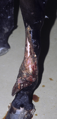
The examination should take place in a clean controlled environment that will allow a thorough assessment, which may require transport to another location. An initial decision should be made whether the wound can be managed with the horse standing or whether general anesthesia will be required. If general anesthesia is likely, immediate care should be provided in the field, and more intensive care and assessment may be provided at the referral center. If the horse is to be referred to another veterinarian, consultation with the referral center is recommended regarding administration of medications and initial treatment in the field. The horse should be adequately restrained to ensure a safe, thorough examination. Use of sedation may be necessary; however, the systemic status of the patient must be considered in cases of severe blood loss or shock. Regional or local analgesia are excellent alternatives to general anesthesia for wounds in the distal portion of the limb. A thorough visual inspection of the wound can provide valuable information about the potential for involvement of nearby associated vital structures and additional considerations that might be required for initial care (Figure 156-1). The specifics of the plan will begin to take shape as progress is made through the process of cleaning, lavaging, and exploring the wound.

Figure 156-1 Severe, contaminated degloving wound over the dorsal aspect of the metatarsus with exposed bone and distal skin flap. Wounds such as this require thorough examination to rule out involvement of the distal tarsal joints, fetlock joint, and digital tendon sheath. The ventral flap should not be sharply transected, but should be retained to aid in wound coverage as contraction occurs. The exposed bone is at risk for sequestrum formation. Debridement may require several days of sequential bandage changes. See Color Plate 17, following p. 704.
Every effort should be made to organize supplies and have them readily available as the examination progresses. An assistant capable of retrieving additional supplies and lending a trained set of hands can be invaluable. The use of examination gloves or sterile surgical gloves is recommended to avoid the potential introduction of iatrogenic infection such as methicillin-resistant Staphylococcus aureus from the veterinarian to the horse. The area surrounding the wound should be prepared first. The wound should be protected from further contamination during the course of these initial preparations. This may be accomplished by filling the wound with sterile water-soluble lubricant, holding a saline-soaked sterile gauze sponge over the wound, or packing the wound with saline-soaked sterile gauze.


