Vitreoretinal surgery

Chapter 12 Vitreoretinal surgery


Introduction


Diseases of the posterior segment, including those affecting the vitreous, retina, choroid, and optic disk, are common in small animals. These diseases may be of congenital, traumatic, inflammatory, degenerative, and neoplastic origin. Diseases of the vitreous affect both cats and dogs, but are more frequent in dogs. Vitreal conditions include persistence of the primary hyaloid vasculature (persistent hyperplastic primary vitreous (PHPV)), vitreal syneresis (liquefaction of the vitreous), asteroid hyalosis (presence of numerous white spherical opacities suspended in the vitreous), vitreal hemorrhage, and vitreal herniation through the pupil. Presentation of vitreous in the pupil and anterior chamber is usually associated with lens displacement and lensectomy, and cataract removal with rupture of the posterior lens capsule and anterior vitreous membrane. Neoplasms, pigment, parasites, foreign bodies, and cysts also affect the vitreous. For these vitreal diseases, surgery may be indicated for diagnosis, or treatment of the vitreal pupillary herniation into the anterior chamber. The removal of diseased vitreous and traction bands in the repair of retinal detachments will be presented in the section on retinal surgeries.


Congenital and degenerative retinopathies are among the more frequently diagnosed clinical disorders in dogs, and the majority of these retinopathies appear inherited. Inflammations of the retina and choroid in dogs are frequent, and often associated with systemic infections. In cats, congenital and degenerative retinopathies are less frequent, but inflammations of the ocular fundus and hypertensive retinopathies are common. Surgeries of the retina and choroid in small animals include retinal or chorioretinal biopsies, and the correction of retinal detachments.


The development of the different types of vitreoretinal surgery in small animals is still early, but these surgeries have been reported since the 1970s. The expensive instrumentation and time-consuming training have delayed application of vitreoretinal surgical procedures in veterinary ophthalmology. Nevertheless, several referral veterinary ophthalmology centers offer this highly specialized service. The inclusion of vitreoretinal surgery in this text will hopefully stimulate its continued development in small animals. In horses, anterior vitrectomy has been recommended in the treatment of recurrent uveitis, and is used widely in Europe.


Surgical anatomy


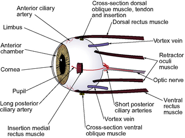
The surgical anatomy of the canine vitreoretinal surgeries includes the extraocular space, insertions of the rectus muscles, and the different landmarks of the external sclera (Fig. 12.1). The surgical approach to the anterior vitreous is usually through the pupil, posterior lens capsule (after extracapsular cataract surgery), and anterior vitreal membrane (after intracapsular cataract and lens removal). Vitreoretinal surgeries involve entry into the vitreal space through multiple small sclerotomies or surgical ports in the pars plana ciliaris. Incisions of the retina are avoided as the resultant hole may produce a rhegmatogenous retinal detachment. Full-thickness retinal holes, whether surgical or spontaneous, require laser, diathermy or cryotherapy to seal the area and stimulate formation of a strong chorioretinal scar. This scar prevents aqueous or vitreous from entering the subretinal space through the retinal break and causing a retinal detachment.


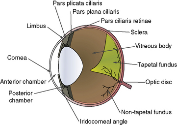
To perform vitreous paracentesis (hyalocentesis), the extent of the pars plana ciliaris or the flat posterior portion of the ciliary body is determined based on measurements posterior to the limbus (Fig. 12.2). The exact anterior and posterior borders of the pars plana ciliaris have been determined in the dog but not in the cat. The width of this tissue varies by quadrant, with the pars plana ciliaris in the lateral quadrant the longest. Hypodermic needle penetration of the ciliary body processes (pars plicata ciliaris) may result in considerable intraocular hemorrhage. Hypodermic needle penetration posterior of the pars plana ciliaris will produce retinal holes. Hence, access to the vitreal space without these serious complications necessitates the external penetration of the sclera and pars plana ciliaris, or the insertion of a hypodermic needle through the pupil after entry into the anterior chamber through a corneal or limbal incision into the anterior chamber.


In order to access the vitreous space during retinal detachment surgery through small scleral incisions (sclerotomies or ports), and for vitreous samples, the critical entry areas are between the end of the anterior pars plicata (ciliary body processes) and the ora ciliaris retinae (beginning of the retina). In a study by Smith and co-workers, the following sites (as determined by calipers posterior to the canine limbus) are recommended for the dog: 1) superotemporal, 6 mm; 2) inferotemporal, 9 mm; 3) superonasal, 5 mm; and 4) inferonasal, 7 mm. Eye size and head size are correlated, and in larger dogs the anterior and middle portions of the pars plana are more posterior. In another study by Sullivan and co-workers, the distances from the limbus to the ora ciliaris retinae were: 1) dorsal quadrant, 9.25 ± 0.81 mm; 2) lateral quadrant, 9.41 ± 0.79 mm; 3) ventral quadrant, 7.18 ± 0.89 mm; and 4) medial quadrant, 5.30 ± 0.77 mm.


Another important site to avoid in the canine is the intrascleral plexus, which drains aqueous humor and is located in the anterior sclera. The intrascleral plexus is located 4–5 mm posterior to the limbus, and may be 4–5 mm wide. Sclerotomies that penetrate the intrascleral plexus often hemorrhage, and judicious cautery is necessary when these sclerotomies are being performed. Under most circumstances, the sclerotomies are posterior to the intrascleral plexus.


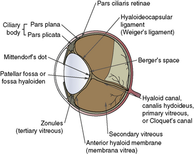
The anatomy of the vitreous is critical to vitreoretinal surgeries, as part of the surgical procedure will nearly always be within the vitreous space (Fig. 12.3). The vitreous humor is normally a clear gel that adjoins the entire retina, and this interface is termed the posterior vitreal membrane. This attachment to the retina appears relatively weak, except at the periphery of the retina (pars plana retinae) and the optic disk. The vitreous border continues anteriorly onto the pars plana ciliaris and, just before the origins and insertions of the zonules or suspensory ligaments between the ciliary body and lens equator, forms the posterior border of the posterior chamber.


The anterior aspect of the vitreous is immediately posterior to the posterior lens capsule, and has been termed the hyaloideocapsular ligament or Weiger’s ligament. The potential space between the posterior lens capsule and hyaloideocapsular ligaments is termed Berger’s space. This separation between the dog’s posterior lens capsule and the hyaloideocapsular ligament appears more potential than real, as tears or penetration of one structure also affects the other. The principal attachment or base of the vitreous appears to be the pars ciliaris retinae or the anterior border of the peripheral retina.


The vitreous body is divided into primary (that vitreous formed and heavily vascularized prenatally) and secondary (that vitreous formed postnatally as the eye grows to normal adult size). The central primary vitreous in adulthood, usually termed Cloquet’s canal, is a clear zone that extends from the optic disk to the central or axial posterior lens capsule. The prenatal blood vessels that traverse this space may persist as remnants at the axial posterior lens capsule (Mittendorf’s dot) or at the optic disk (Bergmeister’s papilla).


The majority of retinal surgeries address repair of retinal detachments. Retinal detachments are microscopic separations of the inner nine layers of the neurosensory retina from the retinal pigment epithelium. Because of retinal development, a potential space exists between the inner nine layers of the retina and the retinal epithelium. With hemorrhage, inflammatory exudates, edema, or the influx of vitreous through a retinal hole, these fluids can separate these retinal layers, and produce the ophthalmoscopic findings of the retinal detachment. Hence, in the repair of retinal detachments, contact must be re-established between the inner nine retinal layers and outer retinal pigment epithelium. Aspiration of subretinal fluids involves the removal of fluids from this space.


Repair of retinal detachments usually includes the placement of a silicone strap (buckle) around the entire globe about the equator, pars plana vitrectomy, retinopexy with laser photocoagulation or cryothermy, tamponade with intravitreal gas or silicone oil, or a combination procedure such as the vitrectomy and a scleral buckle. Hence, knowledge of the insertions of the rectus and oblique extraocular muscles in dogs is important, as these muscles are identified during surgical dissection to place this silicone strap. The bulbar conjunctiva is usually incised 360° posterior to the limbus for exposure of the entire globe.


The sclera constitutes the fibrous tunic for the entire posterior segment. There are several channels or emissaria where the full-thickness sclera is penetrated, and during vitreoretinal surgeries, these regions are avoided (see Fig. 12.1). These channels also represent weak areas of the sclera, and during glaucoma, focal enlargements or staphylomas may develop. At least four vortex veins exit the posterior segment, usually by quadrant and at the equator. These large veins provide the majority of venous drainage from the anterior and posterior segments of the eye and should be avoided. At the 3 and 9 o’clock positions within the external sclera are the long posterior ciliary arteries and veins that are branches of the external ophthalmic artery and vein. These vessels are avoided as they provide the majority of the blood supply to the anterior segment of the eye. Numerous anterior ciliary arteries and veins penetrate the anterior sclera at the insertions of the different rectus muscles. These blood vessels eventually anastomose with other intraocular vessels. Hence, transection of the rectus muscle insertions can decrease the blood supply to the anterior segment of the eye.


Surgical pathophysiology


The pathophysiology of the vitreous and retina are closely related; however, for purposes of discussion, each is presented separately. The vitreous is a clear and fairly firm gel in the young cat and dog, but with aging, the vitreous gradually undergoes liquefaction or syneresis. Intraocular diseases, such as iridocyclitis, the glaucomas, cataract formation, retinal degenerations, and retinochoroiditis accelerate the rate and extent of liquefaction of the vitreous. Hence, if the posterior lens capsule is penetrated during cataract surgery in a young dog, the vitreous is usually a well-formed gel. However, with posterior lens capsule tears in older dogs, the vitreous presentation usually involves a more liquefied vitreous. Vitreous syneresis is also enhanced by cataract and vitreoretinal surgeries.


The role of the vitreous


The appearance of vitreous within the pupil, as after lens displacement or cataract surgery, signals many potential problems. Vitreocorneal contact results in temporary corneal edema. Separation of the vitreous from the cornea may detach the corneal endothelium, resulting in permanent corneal edema. Vitreous can obstruct the pupil and iridocorneal angle resulting in elevations in intraocular pressure (IOP). A miotic pupil can be more easily ‘plugged’ with formed vitreous than a dilated pupil. With vitreous herniated in the anterior chamber and pupil, removal of the vitreous (anterior vitrectomy) is indicated. The vitreous provides variable support to the entire retina in the dog and cat. However, absence of part to all of the vitreous does not automatically result in retinal detachment. Formation of inflammatory traction bands involving the iris, ciliary body, lens capsules, and the vitreous, and the development of holes in the peripheral retina in dogs after cataract and vitreoretinal surgeries, seem more influential in the genesis of retinal detachments than the lack of the vitreous per se. Hence, abnormalities in both the vitreous and retina are key factors in the pathogenesis of rhegmatogenous retinal detachments. Certain breeds of dogs, i.e., Shih Tzu, Boston Terrier, and Toy and Miniature Poodles, develop excessive vitreous degeneration. This degeneration results in considerable syneresis; if the dog is a violent head shaker with toys, rhegmatogenous retinal detachments (RRDs) may result.


Types of vitreoretinal surgery


Vitreoretinal surgeries, presented in subsequent sections, concentrate on vitreous aspiration for diagnosis or injections; vitrectomy (anterior and posterior) for removal of formed or gel vitreous in the pupil and/or anterior chamber, or in treatment of rhegmatogenous retinal detachments; chorioretinal biopsies for tissue analyses; and the surgical management of retinal detachments. Vitreoretinal surgeries are indicated for the rhegmatogenous retinal detachments associated with retinal holes and inflammatory and/or vitreal traction bands.


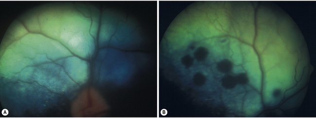
Non-rhegmatogenous retinal detachments are usually treated medically. If possible, the inciting cause is identified and treated. For instance, if systemic hypertension has resulted in the retinal detachment, systemic antihypertensive therapy is administered (Fig. 12.4). If the retinal detachment has resulted from a chorioretinitis, the appropriate therapy is administered. Retinal detachments are also treated symptomatically. Systemic drugs, such as corticosteroids and diuretics, may hasten the reabsorption of the subretinal fluids, and reapposition of the neurosensory retinal layers and the retinal pigment epithelium.


Clinical evaluation of retinal detachments


The broad objective for this type of retinal detachment surgery is the identification of the retinal breaks and sealing these breaks. Detection of retinal breaks in small animals requires meticulous examination of the retina by ophthalmoscopy (usually the indirect method). Mydriasis is necessary to observe as much as possible of the peripheral retina. Often the dog requires sedation to accommodate the time for this examination and the scleral depression. Postoperative capsular and pupillary opacities after cataract surgeries may limit fundic observations. Scleral depression can be achieved under topical anesthesia with a scleral depressor instrument, Jameson muscle hook or a moist cotton swab. The scleral depressor is used to indent the globe a few millimeters posterior to the limbus while simultaneously performing indirect ophthalmoscopy. Most retinal breaks (holes and tears) occur in the peripheral retina.


Once the fundoscopic examination has been performed and any retinal breaks identified and localized, retinal surgery may be indicated. The general principles for the surgical treatment of rhegmatogenous retinal detachments are summarized in Box 12.1, part A. Once the retinal traction is removed and the retinal breaks are sealed, normal physiologic function of the retina may resume with the pigmented epithelium to evacuate the subretinal fluids, and eliminate any space between the neurosensory retina and the retinal pigment epithelial layers.


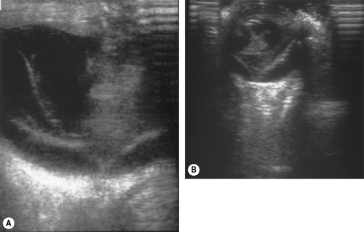
Additional invaluable diagnostic procedures for small animals are electroretinography and ultrasonography. Electroretinography may provide information as to the viability of the retina, and for detecting retinal detachments. Ultrasonography is used particularly to evaluate the posterior segment in which visualization is incomplete because of a small pupil, or lens or vitreal opacities (Fig. 12.5).


Instrumentation for vitreoretinal surgeries


For optimal results in vitreoretinal surgeries, a considerable investment in training and instrumentation is necessary.


Surgical instruments


Basic ophthalmic instruments are necessary to perform the surgical approach for retinal detachments. These instruments are necessary to perform the conjunctival (peritomy) and periocular surgery to isolate and sometimes transect the extraocular muscle insertions. The sclerotomies or small 20 g incisions through the sclera and pars plana of the ciliary body require a basic set of 20 g diameter instrumentation (Box 12.1, part B). These 20 g instruments can be inserted and withdrawn from these ports without causing damage. In addition, several of the instruments are essential. Posterior vitreous cutter, light source, IOP control, and wet-field cautery are necessary. The Machemer lens rests on the cornea and provides irrigation to the cornea, and a wide field of vision and magnification. To inject silicone oil, either a large bore high viscosity cannula with a Luer-lock syringe for manual injection or a special syringe pump and high-viscosity injection, are necessary. A nitrous oxide cryounit with a retinal probe and/or a diode laser with endolaser and indirect delivery modes are essential for retinopexy.


Preoperative considerations for vitreal aspiration and removal


Small animal patients, presented for possible vitreous aspiration, usually have exudative retinal detachments, intense chorioretinitis with vitreal infiltration, and endophthalmitis. Often the ophthalmic disease is part of a systemic infectious disease, and the vitreal aspirate may assist in identification of the infectious agent. Systemic aspergillosis, blastomycosis, and cryptococcosis can often be diagnosed by vitreous aspiration and the demonstration of organisms within the aspirate. During vitreal aspirations, the hypodermic needle can often be directed under direct observation through a dilated pupil to the inflammatory material suspended within the vitreous. Hence, the diagnostic value of vitreal aspirates with a fairly clear anterior segment and lens is quite high. When the cornea, pupil or lens prevents direct observation of the hypodermic needle within the vitreous, the accuracy of the technique declines. Concurrent ultrasonography during vitreous aspiration in eyes with complete opacities can ensure that the hypodermic needle is positioned as accurately as possible within the vitreal body.


Small animal patients with vitreal diseases that may require partial-to-complete surgical excision are usually presented with lens displacement, with or without concurrent secondary and aqueous misdirection (or malignant) glaucoma, or postoperatively after cataract surgery. Formed or gel vitreous, partially herniated in the pupil and anterior chamber, appears as a formed, slightly translucent bulge with occasional fine white fibril strands. Corneal contact will cause focal edema. The pupil margin can be distorted by formed vitreous, and vitreous strands may extend from the pupil into the previous corneal or limbal incision. If the pupil is miotic or miotics are inadvertently instilled, pupillary occlusion with the formed or gel vitreal herniation may result, necessitating additional therapy for the secondary glaucoma.


Medical therapy is usually first attempted for vitreal herniations without corneal contact. Topical corticosteroids and mydriatics are used to suppress the iridocyclitis. The resultant pupillary dilatation decreases the possibility of pupillary occlusion with the herniated vitreous and secondary glaucoma.


Mannitol (1–2 g/kg IV) may be administered once or even twice to dehydrate and shrink the formed vitreous and lower IOP. Ideally, the herniated vitreous will retract behind the dilated pupil and remain. The effects of mannitol in a highly inflamed eye are usually less than optimal because the blood–aqueous barrier is reduced, and the osmotic imbalance caused by the intravascular mannitol is diminished. After 24–72 h of less than successful medical treatment, an anterior vitrectomy is indicated to remove the vitreous from the anterior chamber and pupil.


Preoperative considerations for retinal detachments


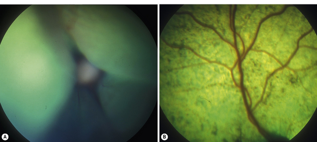
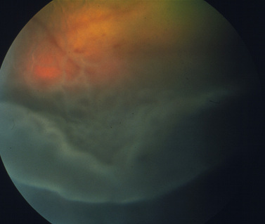
Surgical treatment of retinal detachment has been reported in dogs with serous retinal detachments secondary to optic disk pits or idiopathic (Fig. 12.6), and for rhegmatogenous retinal detachments associated with retinal holes or tears that developed after cataract surgery. As indicated in an earlier section, exudative retinal detachments associated with posterior segment inflammations, systemic hypertension, and other causes are treated by therapy for the specific systemic disorder, and systemic corticosteroids and diuretics to attempt to remove the subretinal fluids before the retinal degeneration becomes advanced. Unfortunately, most retinal detachments are presented late in small animals, and often entire retinas are detached in both eyes. Retinal detachments that develop after lens and cataract removal are often detected earlier during the periodic postoperative examinations (Fig. 12.7).


Surgery of the vitreous


Surgery of the vitreous includes vitreal paracentesis (hyalocentesis), anterior vitrectomy performed through the pupil, and complete vitrectomy performed through the pars plana ciliaris. Vitreal paracentesis is used to obtain liquid vitreous for cytology and culture. Vitreous samples may also be analyzed for drug levels, antibodies, and different substances. In the anterior vitrectomy procedure, often performed at the conclusion of lens removal, vitreous is excised that has entered the anterior chamber or pupil. In the complete vitrectomy procedure, the majority of the vitreous is removed, usually through one or more sclerotomies at the level of the pars plana of the ciliary body.


Modern vitreous surgery techniques have been developed in humans since about 1965. With the demonstration that the excision of vitreous in humans did not result in the detachment of the retina and loss of vision, specialized instrumentation and techniques have been developed in humans to perform vitreoretinal surgeries through small incisions in the pars plana of the ciliary body. Anterior vitrectomy involves the excision of vitreous herniated or protruding through the pupil, as well as pupillary opacities that develop postoperatively after cataract surgeries in small animals. These pupillary opacities usually consist of anterior and posterior lens capsules, organized anterior vitreous and inflammatory fibropupillary membranes. In the former case, the herniated organized anterior vitreous is excised using cellulose sponges or forceps, and vitreous scissors utilizing an anterior vitrector (either portable or attached to a cataract surgical unit). For the postoperative pupillary opacities that develop in dogs and, to a lesser extent, in cats, the anterior vitreous and tough fibrotic membranes require excision by sharp scissors and forceps (anterior vitrectomy and pupillary membranectomy). Depending on its composition, the anterior vitreous may be excised (formed vitreous) or aspirated (liquid vitreous). Sometimes the irregular and often miotic pupil requires coreoplasty (see Chapter 9) to not only excise the opacities within the pupil but also enlarge the pupil to enhance vision.


The instrumentation to perform anterior vitrectomy through a corneal or limbal incision varies, but the basic procedure in small animals may be performed with sharp vitreous scissors, cellulose sponges, cyclodialysis spatula, intraocular forceps, and intraocular scissors. For pars plana vitreoretinal surgeries, a considerable investment in specialized instrumentation is necessary, and only a few veterinary ophthalmology clinical centers have acquired these resources and mastered the surgical techniques. Vitreoretinal surgery, as presented in this chapter, represents one of the surgical frontiers for veterinary ophthalmology where significant advances will undoubtedly occur during the next decade.


Anterior vitrectomy procedures are indicated for excision of persistent hyperplastic vitreous, opacified vitreous, herniation of the vitreous through the pupil, partial excision of the vitreous within the pupil in malignant glaucoma, removal of anterior vitreal foreign bodies, and for excision of vitreous incarcerated within a corneal or limbal wound.


Pars plana vitrectomy is indicated for deeper vitreal opacities, including hemorrhages, treatment of malignant glaucoma, treatment for vitreous prolapsed into the anterior chamber, vitreous removal in endophthalmitis, removal of deep vitreal foreign bodies, excision or transection of vitreous traction and inflammatory bands, and in the management of rhegmatogenous retinal detachments, especially with giant tears.


Hyalocentesis/vitreous paracentesis


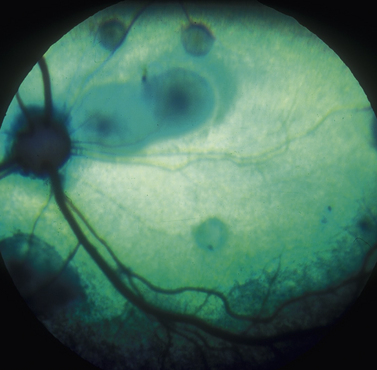
In vitreal paracentesis or hyalocentesis a small quantity of liquid vitreous is aspirated for analysis, usually cytology and bacterial/fungal culture. The formed gel vitreous cannot be aspirated; however, vitreous syneresis or liquefaction occurs with aging, cataract formation, intraocular inflammation, and glaucoma in small animals. As a result, some portion of the vitreous is liquefied, unless the cat or dog is less than 1 year old. Vitreal paracentesis is a most useful diagnostic technique for dogs and cats presented with bilateral exudative retinal detachments or endophthalmitis associated with Blastomyces dermatitidis, Histoplasma capsulatum, Cryptococcus neoformans, Coccidioides immitis, Geotrichum candidum, and Prototheca organisms (Fig. 12.8). Hyalocentesis may also be used to diagnose posterior segment neoplasia; however, this technique is not recommended for this purpose. Direct hypodermic needle contact of the neoplasm is usually associated with hemorrhage. However, if cellular material is present in the vitreous adjacent to the chorioretinal mass, aspiration of the vitreous in this area is reasonably safe.


Vitreous aspiration procedure


Hyalocentesis is performed after the onset of general anesthesia, clipping of the eyelid hair, and cleansing of the eyelid skin, conjunctival and corneal surfaces with 0.5% povidone–iodine solution. The pupil is dilated before the onset of general anesthesia. For convenience, the eyelids are retracted by speculum. The pupil and anterior vitreous are usually visible. The bulbar conjunctiva is grasped by thumb forceps with teeth several millimeters posterior to the dorsal limbus (Fig. 12.9a). By calipers the site to penetrate the bulbar conjunctiva is determined as 6–9 mm (dorsal) to 8–9 mm (lateral) posterior to the limbus. At this position, the 22–23 g hypodermic needle, aimed at the optic disk, should penetrate the sclera and pars plana ciliaris to enter the anterior vitreous (Fig. 12.9b).


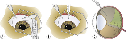
Often once the hypodermic needle is in the anterior vitreous, it can be directly observed. If vitreal opacities or floaters are present, the hypodermic needle is carefully directed to these opacities (Fig. 12.9c). Aspiration of vitreous depends on it being liquefied. Formed vitreous or large opacities may plug the needle and require flushing to relieve the obstruction. The quantity of liquid vitreous aspirated is limited to 0.1–0.25 mL. If larger volumes are removed, an equal volume of saline or lactated Ringer’s solution is injected to immediately restore the lost vitreous volume and IOP.


Hyalocentesis is an excellent diagnostic procedure that can retrieve vitreal opacities for analyses. Identification of the infectious organism, obtained by hyalocentesis, frequently aids in the treatment and prognosis for small animal patients. Treatment after hyalocentesis is usually directed at the pre-existing posterior segment inflammation. Effective treatment of diseases of the anterior vitreous includes the administration of both topical and systemic antibiotics, and/or antifungals. Mydriatics are indicated for the iridocyclitis and are administered at levels sufficient to maintain a reasonably dilated pupil.


Transpupillary vitreal aspirations


Aspiration of liquefied vitreous from the vitreous space may be performed through the pupil during lens and cataract surgery, when formed or gel vitreous herniates through the pupil. Aspiration of some vitreous can be performed in most older dogs during cataract and lens removal surgery, as a portion of the vitreous is usually liquefied. In dogs less than 1 year of age with congenital cataracts, there may be limited-to-no liquefied vitreous, and vitreal aspiration is not possible. Vitreous herniation may follow development of a tear in the posterior lens capsule, or after intracapsular lens and cataract removal. As the posterior lens capsule and anterior vitreal face are in close approximation, damage usually affects both structures.


Appearance of formed vitreous within the pupil may also necessitate excision of any organized vitreous within the anterior chamber with vitreous scissors or the anterior vitrector. Aspiration of liquid vitreous from within the vitreal space is one method to assist in the retraction of formed vitreous behind the pupil.


Part of the pathogenesis of vitreal presentation within the pupil includes not only a tear in the posterior lens capsule and anterior hyaloid face, but also pressure on the vitreal space because of decreased scleral rigidity in dogs and cats under general anesthesia. The possibility of herniation of formed vitreous is reduced by avoidance of tears of the posterior lens capsule during cataract surgery, no instrument pressure on the globe, and the use of neuromuscular blocking agents during general anesthesia in the dog to reduce the extraocular muscle tone on the posterior globe.


Aspiration of liquefied vitreous is usually performed at the completion of phacoemulsification, or extracapsular and intracapsular lens removal. Entry into the anterior chamber has already been established through the small incision used in phacoemulsification, or through the larger corneal or limbal incisions used for extracapsular and intracapsular lens extractions. The cornea is retracted by thumb forceps to expose the anterior chamber and pupil.


The anterior chamber is maintained with viscoelastic agents.


The formed vitreous may be indistinguishable from the aqueous humor or liquefied vitreous, unless some vitreal floaters (cells, fibrils, opacities) are present (Fig. 12.10a). A blunt 18–20 g hypodermic needle, attached to a 1–3 mL syringe, is carefully inserted through the pupil and the remaining posterior lens capsule and/or anterior hyaloid face about 5 mm into the most dorsal vitreal space. Insertion of the hypodermic needle through the external sclera and pars plana ciliaris, as used for hyalocentesis, is not recommended as it may collapse the globe and cause additional formed vitreous to appear in the pupil (Fig. 12.10b). Liquid vitreous is always above the formed or gel portion, and shifts to remain dorsal during the different positions of the eye. As a result, the hypodermic needle to aspirate liquid vitreous must be placed in the upper vitreal body. Liquefied vitreous is slowly aspirated, usually 0.1–0.5 mL. Often with removal of the liquid vitreous, the formed vitreous within the pupil and anterior chamber will partially or completely shift behind the pupil.


Availability of a vitrector has replaced the hypodermic needle approach. During vitrectomy, contact with the iris is avoided, and the tip of the instrument should always be visible. Contact with the iris and ciliary body may result in significant hemorrhage.


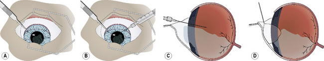
The iridal diaphragm may appear somewhat concave after successful vitreous aspiration, and restoration of all formed vitreous to behind the pupil (Fig. 12.10c). As the gel vitreous and solutions used to lavage the anterior chamber are indistinguishable, a small air bubble may be injected into the anterior chamber to assist in detection of any residual gel vitreous (Fig. 12.10d). The air bubble should be freely maneuverable through the entire anterior chamber when all gel vitreous has been removed or retracted through the pupil. If some gel vitreous remains within the anterior chamber, the air bubble movement is restricted and may outline the formed vitreous. No formed or gel vitreous should remain in the anterior chamber at the conclusion of this technique.


Vitreous aspiration is one of several methods to treat presentation of gel or formed vitreous in the pupil during cataract or lens surgery. Success of this procedure is dependent on some portion of vitreous being liquefied, and is generally unsuccessful in cats and dogs less than 1 year old. This technique may be performed through a tear in the posterior lens capsule and anterior vitreous face. Placement through the pupil must be performed carefully, and a blunt 18–20 g hypodermic needle should be inserted only 5–7 mm posterior to the pupil. Inadvertent needle contact with the retina may produce a retinal hole and predispose to retinal detachment. Nevertheless, this procedure, performed properly, is a useful method to treat vitreal herniation and presentation through the pupil and into the anterior chamber. Medical treatment after vitreal aspiration is directed at the underlying disease, usually postoperative iridocyclitis. The anterior chamber and pupil should be examined daily by slit-lamp biomicroscopy to observe the size and patency of the pupil. Any irregular shape in the pupil may indicate some vitreal contact.


Anterior/partial vitrectomy in small animals


Anterior vitrectomy is divided into two procedures: it may be performed at the conclusion of lens and cataract surgery in the dog and cat, and may also be performed weeks to months after cataract surgery to excise pupillary opacities that also include the lens capsules and anterior vitreous. In the first procedure, which is more common in the dog, anterior vitrectomy is performed when formed vitreous is presented within the pupil, anterior chamber, or the corneal or limbal incision. This complication occurs when the posterior lens capsule and anterior vitreous face are torn or interrupted during phacoemulsification, extracapsular and intracapsular cataract and lens surgeries. Occasionally in intracapsular lens extractions, i.e., anterior lens luxation, formed vitreous may be present in the anterior chamber or even emerge within the corneal or limbal incision as it is performed.


For satisfactory conclusion of cataract and lens removal surgery with vitreal presentation, all formed vitreous must be excised and/or replaced behind the pupil before complete apposition of the corneal or limbal surgical wound.


Vitreous touching the posterior cornea is associated with persistent edema. Vitreous spanning the pupil and incarcerated in the corneal or limbal surgical wound can distort the pupil and serve as the scaffolding for pupillary inflammatory membrane formation. Traction bands from this area may extend into the anterior vitreous and peripheral retina, eventually resulting in retinal detachment. Vitreous within the pupil may also potentially occlude aqueous humor flow, especially when the pupil becomes small.


The anterior vitrectomy procedure is also used for post-cataract pupillary opacification after cataract surgeries in the dog and cat. This procedure may also be combined with coreoplasty (creation of a larger pupil), synechiolysis (cutting or tearing posterior synechia), and removal of extensive fibrotic lens capsules (capsulectomy). These opaque pupillary membranes consist of one or more components including: 1) inflammatory membranes from the postoperative iridocyclitis, when fibrin from the secondary aqueous humor forms the scaffolding for fibroblast proliferation and production of dense collagen fibers; 2) proliferation of remaining lens epithelial cells (Elschnig’s pearls); 3) migration of anterior uveal pigment cells on these membranes from the iris and posterior synechiae; and 4) proliferation of lens fibers as myofibroblasts that contract and distort the posterior lens capsule.


These posterior capsular changes account for the gradual decline in the success rates of cataract surgery in the dog, from 90–95+% at 4–6 weeks postoperatively to about 80% at 2–3 years after surgery. For comparison, posterior lens opacification that requires laser therapy after phacoemulsification cataract surgery in humans occurs in about 50% of the patients within the first postoperative year. Laser intervention in humans is highly successful for restoring visual acuity. In the dog, these capsular changes may also contract the remaining posterior lens capsule, and indirectly (through the zonulary fibers) or directly (by inflammatory traction bands) contribute to the genesis of peripheral retinal holes, tears, and detachments. Part of the rationale for long-term postoperative medical therapy, including topical corticosteroids and systemic non-steroidal anti-inflammatory drugs (NSAIDs), after cataract surgery in the dog is to either retard or prevent formation of these pupillary membranes. The cat, in contrast to the dog, shows a less intense iridocyclitis after cataract surgery, and a lower tendency to develop these pupillary membranes.


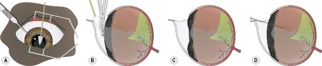
When the anterior vitrectomy is performed immediately after lens or cataract removal, the majority of the procedure is performed within the anterior chamber. The formed vitreous may be clear or contain suspended cells, fibrin, vitreous opacities (such as with hyalosis), and blood. In the anterior vitrectomy procedure for anterior chamber vitreal presentation, small cellulose surgical sponges are touched to the formed vitreous (Fig. 12.11a).The adherent gel vitreous is slowly retracted and carefully cut with sharp iris or vitreous scissors. The scissors are held parallel to the surface of the iris to minimize traction on the deeper vitreous and indirectly on the retina (Fig. 12.11b). All gel vitreous within the anterior chamber may be removed by this procedure. The iris surface should be slightly concave afterwards because of the loss of formed vitreous immediately posterior to it (Fig. 12.11c). Many phacoemulsification units also offer vitrectomy capacity that can be used to cut and aspirate the formed vitreous from the anterior chamber.


Stay updated, free articles. Join our Telegram channel

Oct 14, 2016 | Posted by in SUGERY, ORTHOPEDICS & ANESTHESIA | Comments Off on Vitreoretinal surgery

Full access? Get Clinical Tree

Get Clinical Tree app for offline access