Viral Infections

Chapter 111 Viral Infections


INTRODUCTION


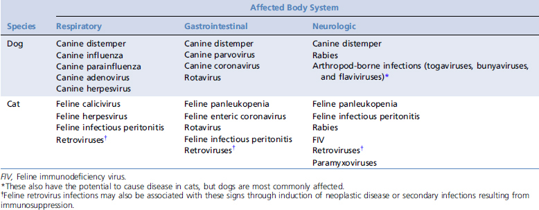
A large number of viruses may cause acute and severe illness in dogs and cats (Table 111-1). The most common or important viral infections that may come to the attention of emergency and critical care veterinarians are canine parvovirus (CPV), canine distemper virus (CDV), canine influenza virus, feline panleukopenia virus, feline herpesvirus (FHV-1), feline calicivirus (FCV), feline infectious peritonitis virus (FIPV), feline immunodeficiency virus (FIV), feline leukemia virus (FeLV), and rabies virus infection. The FIV and FeLV status of all cats should be determined on arrival by questioning the owner or testing using in-house enzyme-linked immunosorbent assays for FeLV antigen and FIV antibody. Because these infections may be detected in asymptomatic cats, and because some cats may eliminate FeLV, positive test results alone are not reason for euthanasia. CPV infection is covered in the following chapter. Other viral diseases that may present to emergency and critical care veterinarians include enteric viral infections such as rotavirus and coronavirus infections, feline paramyxovirus infection, pseudorabies virus infection, vector-borne viral infections such as West Nile virus infection, infectious canine viral hepatitis, and canine herpesvirus infection.


An extensive discussion of the etiology, clinical signs, diagnosis, treatment, and prevention of every one of these infections is beyond the scope of this chapter. Instead, the purpose of this chapter is to provide the reader with an update on selected common and important viral infections in dogs and cats that may present to emergency and critical care veterinarians. Treatment of viral infections is largely supportive and symptomatic and usually includes intravenous fluid therapy, early enteral or parenteral nutrition, antiemetics, analgesia, and oxygen therapy when pulmonary disease is present. Blood products may be needed for cats with retroviral infections. Antibiotics may be needed for secondary bacterial infections. Attempts to culture secondary bacterial invaders and determine sensitivity to antimicrobial agents should be considered before commencing antimicrobial therapy. Use of antiviral medications is still limited in dogs and cats, and controlled studies are lacking.


CANINE DISTEMPER VIRUS INFECTION


CDV infection is a contagious disease of dogs that may involve the gastrointestinal (GI), respiratory, or neurologic systems. Distemper still occurs sporadically, even in vaccinated dog populations. Disease most commonly occurs in dogs 3 to 6 months of age, when maternal antibody level is declining, but can occur in older dogs that have been vaccinated infrequently, especially following stress, immunosuppression, or contact with other affected dogs.1


CDV is an enveloped ribonucleic acid (RNA) virus that belongs to the family Paramyxoviridae. The virus survives for about 3 hours at room temperature and is highly susceptible to routine hospital disinfectants such as quaternary ammonium compounds. Several strains of CDV exist and vary in pathogenicity. Some, such as the Snyder Hill strain, are more likely to produce neurologic disease than others. A study has documented the existence of CDV strains that differ from vaccine strains and those previously documented in the United States.2


CDV is shed in respiratory secretions for up to 90 days after infection. Initial replication of CDV is in lymphoid tissue, and viral destruction of lymphocytes results in lymphopenia and pyrexia. Approximately 1 week after infection the virus spreads to epithelial tissues (lungs, GI tract, kidney, bladder) and the central nervous system (CNS), and virus shedding begins. Poor cell-mediated immunity (CMI) is associated with spread of the virus to a variety of tissues, severe respiratory and GI signs with or without CNS involvement, and death. Dogs with an intermediate or delayed CMI response may develop persistent infection of the uvea, CNS, and footpad and nasal epithelium, leading to neurologic, cutaneous (hard pad), and ocular signs such as chorioretinitis. Infection with CDV is highly immunosuppressive, and secondary infections with opportunistic organisms such as Toxoplasma and Salmonella may occur.


Distemper should be high on the list of differential diagnoses for any dog with respiratory and/or CNS signs. Mild signs are common and resemble those of kennel cough. Severe, generalized distemper may begin with a serous to mucopurulent conjunctivitis and rhinitis, and progress to include signs of lower respiratory disease, depression, anorexia, vomiting and diarrhea, severe dehydration, and death. Neurologic signs then occur in some dogs, either with systemic illness or after a several-week delay. Neurologic signs are frequently progressive despite treatment and are a poor prognostic sign. Myoclonus, an involuntary twitching of various muscle groups, can be most pronounced when affected dogs are resting and is virtually pathognomonic for CDV infection. Ocular signs may consist of sudden blindness due to optic neuritis, chorioretinitis, or retinal detachment. Cutaneous signs may be useful for prognostication. Footpad and nasal hyperkeratosis often are accompanied by neurologic complications, whereas the presence of vesicular and pustular dermatitis implies a good CMI response and rarely is associated with neurologic complications.


Physical examination of dogs suspected to have distemper should include a fundic examination, careful inspection of the skin, including the nose and footpads, and careful thoracic auscultation. Any dog suspected to have distemper should be placed in isolation if possible. This may be complicated by a requirement for oxygen therapy.


The most commonly used diagnostic test for distemper is cytologic examination of conjunctival scrapings. Acutely, these may show cytoplasmic inclusions in epithelial cells when stained with Wright or Diff-Quik stain (Color Plate 111-1). The sensitivity of cytology is increased following application of immunofluorescent antibody to smears by regional diagnostic laboratories. Smears should be air dried and, if possible, fixed in acetone for 5 minutes before transport. Intracytoplasmic inclusions may also be seen in erythrocytes, lymphocytes, other white blood cells, and cells within the cerebrospinal fluid (CSF). Thoracic radiography may reveal an interstitial pattern, or an alveolar pattern with secondary bacterial bronchopneumonia. Analysis of CSF may show increased protein and cell count, and measurement of anti-CDV antibody in the CSF can also be useful for diagnosis in dogs with neurologic signs. Other antemortem diagnostic tests for distemper include immunohistochemistry for CDV antigen on biopsies of nasal mucosa, footpad epithelium, and haired skin of the dorsal neck, and reverse transcriptase–polymerase chain reaction (RT-PCR) testing for viral nucleic acid. Samples suitable for RT-PCR testing include buffy coat cells, whole blood, serum, CSF, and urine.3 With any PCR assay, quality control can be problematic, and the laboratory should be consulted to ensure adequate positive and negative controls are included. Virus isolation is difficult and is not widely used for diagnosis.


Modified live vaccines can prevent canine distemper and should provide at least partial protection even in the face of variant strains. The interested reader is referred to a comprehensive review of vaccination for CDV for further information on the topic.1

Stay updated, free articles. Join our Telegram channel

Sep 10, 2016 | Posted by in SMALL ANIMAL | Comments Off on Viral Infections

Full access? Get Clinical Tree

Get Clinical Tree app for offline access