Allergic Airway Disease in Dogs and Cats and Feline Bronchopulmonary Disease

Chapter 20 Allergic Airway Disease In Dogs And Cats And Feline Bronchopulmonary Disease


Definition of Allergic Airway Disease


Allergic airway disease in dogs and cats encompasses a broad spectrum of diseases that are somewhat poorly defined, but the clinical signs and pathologic appearances are similar regardless of causes. Diseases commonly included in this category are parasitic allergic airway disease, allergic bronchitis (eosinophilic bronchopneumopathy), feline asthma, and pulmonary infiltrates with eosinophils (PIEs). These diseases typically are characterized by bronchial or alveolar inflammatory changes including submucosal wall edema, increased bronchial secretions, smooth muscle hypertrophy, and smooth muscle constriction of the bronchioles and small bronchi. Histologically, there is typically a predominance of eosinophils within the airways and submucosa of the bronchial tree. Clinical signs include labored breathing, rapid shallow breathing, increased expiratory effort, and cough. Studies have shown variable degrees of inherent hypersensitivity in the bronchiolar smooth muscle in small animals with lower airway disease.1,2 Animals may have an acute onset of respiratory signs with completely reversible changes or may develop chronic disease (defined as more than 2 months duration) that is associated with irreversible bronchial wall alterations.3 The pathogenesis of these diseases has not been as thoroughly investigated as has human asthma. One should avoid terming small animal allergic airway disease as asthma because the pathogenesis and definition are much less clear than they are in humans.4


PATHOGENESIS OF SMALL ANIMAL ALLERGIC RESPIRATORY DISEASE


The pathophysiology of small animal allergic airway disease is less well understood than human asthma, but it is clear that these diseases in small animals are characterized clinically by a cough, typically a prominent increase in expiratory effort with or without appreciable wheezes, and a predictable response to glucocorticoids.2,4,9 Allergic airway disease in small animals commonly causes an increase in the numbers of eosinophils within the airways, hyperinflation of the lungs, and thickening of the bronchi and bronchioles. Lower airway inflammation, in response to either an extrinsic noxious stimulus or intrinsic hypersensitivity to antigenic stimulation, is a known component in animals with allergic respiratory disease. The airway inflammation causes mucosal edema, airway smooth muscle hypertrophy and constriction, and excessive production of airway secretions.3,9 Although similar in clinical picture and treatment, diseases such as feline and canine bronchitis should not be termed allergic in nature because they do not fit all of the above criteria (particularly overabundance of eosinophils in the airways). Diseases that should be included in small animal allergic airway disease include canine allergic bronchitis (also termed eosinophilic bronchopneumopathy), parasitic larval migration, PIE, and feline asthma.10 Although the clinical picture and treatment of feline bronchopulmonary disease and feline asthma are similar, it is important to remember that they may have separate causes, because the cytopathologic features differ.


PARASITIC ALLERGIC AIRWAY DISEASE


Intestinal parasite migration as well as primary pulmonary parasitism can cause a parenchymal or lower airway allergic inflammatory response. The most common migratory parasite to cause an allergic response in the canine lungs is Toxocara canis. An inflammatory “allergic” reaction can take place in the lower airways and parenchyma of young dogs when this parasite migrates through the lungs as part of its normal development. Because of antigenic stimulation and the eosinophilic infiltrate induced by the larvae, these dogs may develop signs of respiratory disease that can vary in intensity.11


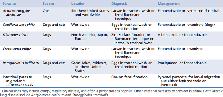
Other, less common parasites known to migrate through the lungs include Ancylostoma caninum (dogs only) and Strongyloides stercoralis (dogs or cats). Primary lung parasites include Paragonimus kellicotti, Aelurostrongylus abstrusus, Capillaria aerophila, and Filaroides hirthi (Table 20-1). Dirofilaria immitis (heartworm infection) can also cause an allergic inflammatory response when large numbers of antimicrofilarial antibodies entrap microfilariae within the pulmonary capillaries.12 All of these parasites elicit predominantly a type I hypersensitivity reaction in the lungs that leads to bronchoconstriction and inflammation within the airways and lung parenchyma.11


Clinical signs associated with larval migration or primary pulmonary parasitic infection vary markedly from asymptomatic to severe coughing, wheezing, and respiratory distress. A complete blood count may show eosinophilia or basophilia, however this finding is not always present in animals suffering from parasitic allergic airway disease. Chest radiographs can show a variety of changes, including interstitial infiltrates, bronchial thickening, and even alveolar consolidation. Ancylostoma caninum and Toxocara canis can be seen using routine fecal flotation techniques. 10Strongyloides stercoralis is more reliably found with the Baermann technique. However, negative fecal examination results do not rule out the possibility of migrating larval airway disease. Ova are often difficult to find on fecal examination because larvae typically begin to migrate through the lungs before shedding ova into the intestinal tract.13


Initially, a course of an appropriate antihelminthic medication (ivermectin or fenbendazole) can be used for treatment, particularly in mild to moderate clinical cases (see Table 20-1). Appropriate treatment for infection with D. immitis is discussed elsewhere.14 In situations in which the clinical signs are severe, or fail to resolve completely, an antiinflammatory dosage of prednisone (0.5 to 1 mg/kg q24h) may be used to help control the disease manifestations.10,11


CANINE ALLERGIC BRONCHITIS OR EOSINOPHILIC BRONCHOPNEUMOPATHY


Canine allergic bronchitis (eosinophilic bronchopneumopathy) is characterized by pulmonary hypersensitivity with eosinophilic infiltration of lung and bronchial mucosa. The signalment of dogs with this disease tends to be different from that of either PIE or canine chronic bronchitis; these dogs tend to be younger (mean ± SD = 3.3 ± 2 years) and Siberian Huskies and Alaskan Malamutes are overrepresented. These dogs usually are in good physical condition, but show clinical signs such as coughing, labored breathing, or nasal discharge that is mucopurulent or yellow-green in appearance.12,15


The most common radiographic finding in dogs with canine allergic bronchitis (eosinophilic bronchopneumopathy) is a diffuse, prominent, bronchointerstitial pattern. Forty percent of dogs have alveolar infiltrates (due to secondary pneumonia in some cases), and 26% have radiographic signs of bronchiectasis. A peripheral eosinophilia is present in about 60% of cases. Bronchoscopy typically reveals abundant yellow-green mucus or mucopurulent material, thickening with irregularities or polypoid changes to the mucosa, and exaggerated closure of the airways during expiration. Cytologic findings in fluid obtained from a bronchoalveolar lavage (BAL) or endotracheal wash (ETW) include more than 50% eosinophils in 87% of dogs and between 20% and 50% eosinophils in 13% of dogs.15


The mainstay of treatment for animals with this disease is glucocorticoids, with an induction dosage of prednisone of approximately 1 mg/kg q12h, although larger dogs often require lower dosages. Most dogs will relapse within months of discontinuing the steroids, but some dogs may remain disease free for years. A maintenance dosage of prednisone (0.25 to 0.5 mg/kg q48h) is suggested in an attempt to maintain remission. Other immunosuppressive drugs and inhaled medications have not been evaluated objectively for their efficacy in treating animals with this disease. Culture and sensitivity testing should be performed on the BAL or ETW fluid in order to rule out a secondary pneumonia. It is important to stress to the owner that this disease requires life-long management and there may be unwanted side effects from the long-term use of steroids.12,15

Stay updated, free articles. Join our Telegram channel

Sep 10, 2016 | Posted by in SMALL ANIMAL | Comments Off on Allergic Airway Disease in Dogs and Cats and Feline Bronchopulmonary Disease

Full access? Get Clinical Tree

Get Clinical Tree app for offline access