Pulmonary Contusions and Hemorrhage

Chapter 25 Pulmonary Contusions and Hemorrhage


INTRODUCTION


Pulmonary contusions consist of pulmonary interstitial and alveolar hemorrhage and edema associated with blunt chest trauma, usually after a compression-decompression injury of the thoracic cage. Such injury is most commonly associated with motor vehicle trauma1 and high-rise falls2 in cats in urban areas. Other causes include animal interactions (e.g., horse kicks), human abuse, and shock waves from explosions.


Thoracic trauma has been reported in 34%,3 38.9%,4 and 57%5 of dogs and 17% of cats4 that sustained limb fractures in road traffic accidents. Pulmonary contusions were the most prevalent lesion in roughly 50% of animals with thoracic injuries, although patients may display few clinical signs associated with the contusions. They may occur as an isolated abnormality or in combination with other thoracic injuries including pneumothorax, pleural effusion, rib fractures, diaphragmatic rupture, cardiac arrhythmias, and pericardial effusion.4,6 Pulmonary contusions may be the most significant abnormality present; in one study, only 32% of dogs had concurrent fractures or luxations.7 It is worth noting that in another study 79% of the dogs with abnormal thoracic radiographic findings or low arterial partial pressure of oxygen (PaO2) had no physical findings that were suggestive of thoracic injury on initial examination.5


The clinical manifestations of pulmonary contusions, as in any thoracic trauma, can be acute and lead to immediate, severe respiratory distress, or may develop progressively over several hours after the injury. Radiographic changes may also be delayed. Because aggressive fluid therapy and general anesthesia have the potential to worsen contusions, the emergency clinician must not discount the possibility of their presence when evaluating more dramatic injuries, even if clinical signs of thoracic injury or respiratory distress are not apparent initially.


PATHOPHYSIOLOGY AND PATHOLOGY


Pulmonary contusions result from the release of direct or indirect energy within the lung. High-velocity missiles and blasts also lead to pulmonary contusions as shock waves pass through the parenchymal tissue and lead to bleeding into the alveolar spaces and disruption of normal lung structure and function. Several mechanisms have been postulated as important in the etiology of pulmonary contusion.8


Due to the compressible nature of the thoracic cage, acute compression and subsequent expansion lead to transmission of mechanical forces and energy to the pulmonary parenchyma. As a result, the lung is injured directly by the increased pressure in the so-called spalling effect, a shearing or bursting phenomenon that occurs at gas-liquid interfaces and that may disrupt the alveolus at the point of initial contact with shock waves. The “inertial effect” that occurs when low-density alveolar tissue is stripped from heavier hilar structures as they accelerate at different rates results in both mechanical tearing and laceration of the lungs. Finally, an “implosion effect,” resulting from rebound or overexpansion of gas bubbles after a pressure wave passes, can lead to tearing of the pulmonary parenchyma from excess distention.9,10 The parenchyma may also be injured by the displacement of fractured ribs. Subsequent hemorrhage results in bronchospasm, increased mucus production, and alveolar collapse due to decreased production of surfactant.11


Damage to the lung leads to complex changes in respiratory function. The parenchymal damage causes ventilation-perfusion mismatch as the alveoli are flooded with blood and underventilated. There is also an increase in lung water due to the accumulation of protein-rich edema, subsequently decreasing lung compliance.12 However, the initial vasoconstriction in response to local hypoxia (hypoxic pulmonary vasoconstriction) may be followed by a further decrease in local perfusion secondary to vascular congestion and thrombosis. This results in reduced perfusion to the unventilated lung, thus reducing the shunt fraction. The patient subsequently displays dyspnea from hypoxemia. Either hypocarbia or hypercarbia may be present depending on the severity of the contusions and the effects of concurrent injuries on ventilation.


In animals that survive the initial hours, the respiratory derangements associated with pulmonary contusions usually resolve in 3 to 7 days, but delayed deterioration may occur. Delayed clinical signs of pulmonary dysfunction may occur from complications such as bacterial pneumonia or acute respiratory distress syndrome (ARDS) secondary to the local or systemic inflammatory response.11 The frequency of these complications has not been well described in dogs and cats. In humans, pulmonary contusions cause severe immunodysfunction both locally and systemically, and this immunosuppression is associated with a decreased survival rate if a septic insult occurs.13


Histologic progression of pulmonary contusions has been demonstrated in a canine experimental model.14 Immediate interstitial hemorrhage is followed by interstitial edema and infiltration of monocytes and neutrophils during the first few hours. Twenty-four hours after injury, the alveoli and smaller airways have been filled with protein, red blood cells, and inflammatory cells. At this stage, the normal architecture has been lost and edema is severe. Alveoli adjacent to the affected region remain normally perfused, but they are less compliant due to the edema and disruption of the surfactant layer. Thus, they are poorly ventilated, which leads to an increase in ventilation-perfusion mismatch.12,15-17 Furthermore, experimental studies in pigs have demonstrated that local pulmonary contusions may lead to generalized pulmonary dysfunction secondary to impaired surfactant activity and a subsequent decrease in alveolar diameter.18 Forty-eight hours after injury, healing has started and the lymphatic vessels are dilated and filled with protein. The parenchyma and affected airways contain fibrin, cellular debris, granules from type II alveolar cells, neutrophils, and macrophages.12 Another study found that within 7 to 10 days post trauma, canine lungs were almost completely healed with little scarring.16


DIAGNOSIS


Imaging: Radiology and Computed Tomography


Dyspneic patients should undergo stabilization before imaging is attempted. Animals that sustain thoracic trauma may have multiple thoracic injuries, making a precise diagnosis based on the physical examination alone challenging. Imaging studies may be helpful in identifying the injuries. However, as with all dyspneic patients, the risk-to-benefit ratio of the imaging procedure should be considered carefully.


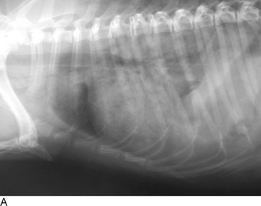
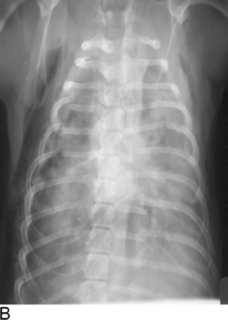
Radiographic changes in patients with pulmonary contusions consist of areas of patchy or diffuse interstitial or alveolar lung infiltrates that can be either localized or generalized (Figure 25-1). Radiographic changes may lag behind clinical signs by 12 to 24 hours and therefore “normal” radiographic findings may be seen in animals with pulmonary contusions. Patients with more severe radiographic changes initially may require a longer duration of oxygen supplementation and longer hospitalization times. However, the relationship between severity of the contusion based on radiographic changes and survival has not been established.7


Stay updated, free articles. Join our Telegram channel

Sep 10, 2016 | Posted by in SMALL ANIMAL | Comments Off on Pulmonary Contusions and Hemorrhage

Full access? Get Clinical Tree

Get Clinical Tree app for offline access