5 Viral diseases
There are important principles associated with confirmation of diagnosis in many of the viral conditions. The collection of samples for viral identification by electron microscopy or for culture usually requires special procedures because the viruses may be relatively fragile, being easily affected by minor changes in pH, light exposure, disinfectants etc., and in any case may only be found in significant numbers in localized sites within the lesion or in ‘early’ lesions. Modern PCR and antigen ELISA tests can be very sensitive at detecting the presence of any virus particles (Martens et al 2001). Also serological evidence can sometimes be used where a positive viral detection is not possible; this is particularly useful for systemic viral conditions with cutaneous signs. Additionally, skin lesions caused by viral diseases may be confusingly contaminated or complicated by bacterial or fungal infections.
Probably the most important primary viral skin diseases are those caused by pox viruses or papillomaviruses. However, there is little specific information about most of these viruses. Vesicular stomatitis is an important disease because it is probably transmissible to other animals and possibly even to humans, and in ruminants and pigs the condition can resemble foot and mouth disease; it is reportable in most countries but it can easily pass unnoticed because the signs are usually transient and can be mild. There are also several other important diseases with cutaneous signs such as African horse sickness, rabies (see pruritus, p. 442) and viral arteritis. In most of these conditions the dermatological symptoms are themselves trivial compared to the main ones but they can provide useful diagnostic support.
Horse pox (vaccinia)
Profile
There is a potential zoonotic implication.
 Key points: Horse pox (vaccinia)
1. Rare disease probably related to a specific but unidentified equine pox virus or vaccinia (the cowpox-derived attenuatedvirus used for human smallpox vaccination).
2. Localized forms are more likely and the muzzle-mouth is the commonest site. Pastern dermatitis can be present.
3. Diagnosis can be confirmed by electron microscopy and by PCR methods.
4. Lesions can be painful but are usually transient; early and mild cases are seldom reported to veterinarians.
5. There is no treatment and no vaccination. Immunity following infection is probably life-long.
Clinical signs
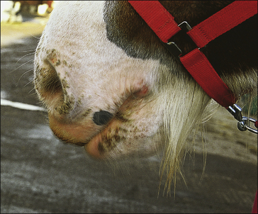
The disease occurs in two forms. In the buccal form, pox-like lesions occur in the mouth and skin of the lips and nostrils (buccal horse pox/contagious pustular stomatitis) (Fig. 5.1). This form lasts for around 3 weeks and is not considered to be serious. In some cases it may be overlooked completely. A condition resembling this condition was reported in a donkey (Jayo et al 1986).
Vesicles rapidly develop over a period of about 4 days and burst to become dark/black umbilicated ulcers and then pustules with surface crusts and exudate. The lesions may be painful to the touch but are not usually pruritic. This form may closely resemble molluscum contagiosum (see p. 133) and in fact it may be the same disease.
Mild systemic signs (pyrexia and depression) may be seen.
Lesions may be detected on humans in contact with affected horses.
Differential diagnosis
• Molluscum contagiosum: geographically largely restricted to South Africa.
• Papillomatosis: very common, easily recognized, spontaneously resolving condition of young horses at grass in particular.
• Immune-mediated disease, such as pemphigus complex: generalized signs are more usual.
• Viral papular dermatitis: rare, slowly resolving viral disease – can be very similar.
• Dermatophytosis (ringworm): characteristic epidemiology and size and shape of lesions – diagnostic mycology.
• Dermatophilosis: this is more exudative and occurs commonly; there is characteristic bacterial identification.
• Coital exanthema: a transient, venereal infection that usually affects only the external genitalia.
• Sarcoid: long-standing/persistent neoplastic condition with multiple clinical manifestations.
Diagnostic confirmation
• Biopsy may show intracytoplasmic inclusion and ‘ballooning’ and sometimes microvesicle formation in keratinocytes in the stratum spinosum.
• Immunohistochemistry using indirect or direct fluorescent antibody stains on normal sections of affected skin.
• Virus isolation is characteristic and PCR methods can be confirmatory.
• Electron microscopic detection of the characteristic ‘brick-shaped’ virus particles.
Molluscum contagiosum
Profile
This is a mildly contagious cutaneous infection of horses of any age caused by an unclassified pox virus closely related to the horse pox group. Restricted geographically at present to South Africa but some cases may be encountered in other places including southern Europe (Scott & Miller 2003). Horse-to-horse transmission occurs slowly even when there is relatively close contact between infected and non-infected individuals. Spread by fomites is possible but less likely. A similar disease occurs in humans and the virus and the pathology are very similar (if not identical). Horses may be infected by humans (Thompson et al 1998).
 Key points: Molluscum contagiosum
1. Rare geographically restricted, largely to areas of southern Africa, viral (molluscipox) skin disorder affecting horses and humans.
2. Possibly transmitted to horses from humans or by direct close contact between horses or by insect vectors.
3. Miliary, transient or long-lived papular lesions with a waxy appearance occurring all over the body with most in the hairless skin such as the inguinal region, and face.
4. Diagnosis is easily confirmed by characteristic histopathology.
5. No treatment is warranted or available. Slow resolution may occur but some lesions may persist.
Clinical signs
The condition is non-painful and non-pruritic. There are no apparent systemic effects.
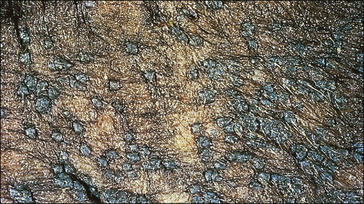
Multiple, raised, well-defined grey/white papules with a waxy surface (1–6 mm) occur primarily in hairless or sparsely haired (glabrous) areas such as the penis, prepuce, scrotum, mammary glands, thighs (Fig. 5.2), axilla and muzzle. Other body areas such as the rump, neck and chest can also be affected and focal alopecia is a common sign (Fig. CD5 • 1A and B) . Individual lesions may coalesce and form larger aggregations closely resembling papillomas (see Fig. 5.6) (Moens & Kombe 1988).
Differential diagnosis
• Papillomatosis: transient fibro-papillomatous condition with a characteristic pattern of development in many parts of the world. Differentiation is difficult where both conditions are endemic; histology and electron microscopy is characteristic.
• Uasin Gishu disease/viral papular dermatosis: can be very similar in appearance but occurs in geographically distinct regions.
• Immune-mediated skin diseases (pemphigus complex/sarcoidosis, etc.): multifocal seborrheoic persistent autoimmune dermatosis.
• Occult/verrucose sarcoid: characteristic variations in clinical appearance and persistence with a tendency to exacerbation on interference.
• Dermatophytosis (ringworm): transient, alopecic and scaling dermatosis with characteristic epidemiology and mycological examinations.
• Horse pox: localized, transient and geographically distinct regions.
• Coital exanthema: transient venereal pustular condition restricted largely to external genitalia.
Vesicular stomatitis (red nose; sore nose)
Profile
This is a sporadic contagious viral disease of limited geographical distribution caused by Lyssa virus (family Rhabdoviridae). Most cases are encountered in spring and late summer. The virus is probably spread by insect vectors (biting, sucking and surface-feeding flies including Culicoides spp., Simulium spp. and muscid flies) and possibly by fomites (Knight & Messer 1983). The prevalence is highest in closely congregated animals.
Clinical signs
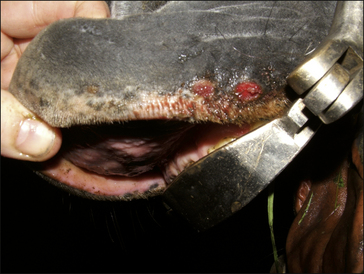
Inflammation and vesicle a lesser extent on the skin of the nose, mammary gland and prepuce (Fig. 5.3 and Fig. CD5 • 3A–C ). Coronet and digital skin are also sometimes affected. Vesicles rupture leaving a very painful, small, ulcerated lesion.
Scars of previous infection on the skin may be seen as focal depigmented spots (Donaldson & Ferris 1995).
 Key points: Vesicular stomatitis
1. Geographically largely restricted to North and South America.
2. Probably most significant transmission is by biting insects but direct close contact is also a possible route. Feeding troughs and water buckets may be involved in the spread between horses.
3. The transient signs are usually restricted to the mouth and lips but are occasionally found elsewhere.
4. Miliary, small, painful vesicles and ulcers cause inappetence and salivation (often with some blood). Lesions on the coronary band can lead to lameness.
5. Treatment is not warranted – spontaneous resolution is the norm.
Differential diagnosis
• Viral papular dermatosis: an unusual viral disease with transient unilateral papules.
• Molluscum contagiosum: a geographically restricted viral wart-like disorder showing waxy papules over extensive areas; resolution is usually very slow.
• Horse pox (‘mouth pox’ forms): transient vesicular and papular lesions mainly around the mouth and nose.
• Bullous pemphigoid/pemphigus vulgaris: a multifocal or diffuse (generalized) exfoliative autoimmune dermatopathy with characteristic histological findings.
• Contact irritants/chemical injuries: history of exposure is usually established and some can be detected directly.


 . There is usually little or no dermal inflammation.


 Key points: Equine viral arteritis


