Chapter 16 Veterinary Participation in Puerto Rican Crested Toad Program
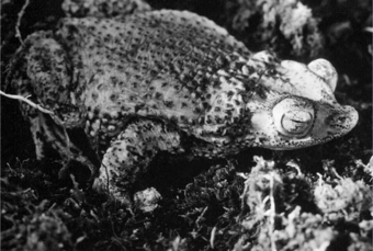
The crested toad, or sapo concho (Bufo [Peltophryne] lemur), is one of eight endemic West Indian bufonids and is now found only on Puerto Rico. It has prominent supraorbital crests and an upturned snout. The toads are sexually dimorphic; females are 80 to 120 mm long and ash gray–white to charcoal black; whereas the males are slightly smaller, 70 to 80 mm long, with a brighter, yellow-green coloration (Figure 16-1).
CONSERVATION
The PRCT had not been seen for more than 35 years until its rediscovery in 1965.5 No record of captive animals exists before 1980. Puerto Rico is a mountainous island, and only two isolated toad populations were known to exist: a northern population, now believed to be extirpated, and a southern population believed to be as low as 1500 to 2000 in the 1980s.6 These populations have been isolated since the Pleistocene, and they are managed in captivity as two genetically distinct groups. Because of its fossorial nature, the species is difficult to inventory. During the day, toads live underground, usually entering secure, moist crevices or holes in the limestone karst. The largest known breeding population is found in Guanica Forest Reserve on the southwestern coast.
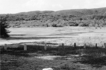
Typically, when the rainfall exceeds 18 cm over a 24-hour period, the adults emerge to breed at temporary ponds near the coastal beach. The breeding area is located adjacent to the sea, and inundation by seawater during hurricanes is a real threat. Before 1984 the breeding pond was drained to provide easier beach access. When this practice was stopped, it was discovered that toads were using this pond as a breeding site (Figure 16-2). The northern toad populations, found near Quebradillas, had bred in concrete walk-in cattle troughs together with B. marinus. Here, no more than 25 PRCTs had been seen at any one breeding episode. There has been no sighting of northern toads since 1992, and wild northern toads at this time are functionally extirpated. The remaining northern toads that now exist in zoos originate from a single breeding of the last pair of this race. In May 2006, tadpole descendants from these were released in newly constructed ponds near Arecibo, the first time in 15 years.

Fig 16-2 Breeding habitat in the Guanica Forest Reserve, Puerto Rico, in the dry season.
(Courtesy B. Johnson.)
For several years, censuses in the Guanica Forest showed a population at the breeding pond of fewer than 300 toads, but in 2004 more than 600 adult toads appeared at the pond in three separate breeding events.1 In 2005, four breeding events took place involving 2200 toads.
The PRCT species was considered suitable for a species survival plan (SSP) under the auspices of the Association of Zoos and Aquariums (AZA) in 1984 and was the first amphibian included in the SSP program. The 21 facilities currently holding 340 Bufo lemur manage approximately 400 captive animals as one population. Since the late 1980s, a captive breeding and release program has been undertaken to augment the wild population. Releases are only considered for suitable habitats beyond the range of the extant population. To date (2006), approximately 100,000 tadpoles hatched in North American zoos have been released in Puerto Rico.7 Additional breeding sites have been constructed, and in 2003, for the first time, adult toads grown up from captive-bred tadpoles were breeding at the new sites.
Reproduction
Several chemical analogs of LHRH exist, and not all work well in each species. The analog most often used for anurans is des-Gly (d-Ala) LHRH ethylamide (catalog #L4513, Sigma, St. Louis). To achieve natural reproduction, it is necessary not only to induce gamete production and release, but also to promote behavioral amplexus for fertilization of released ova. Experiments have shown that hCG injected into PRCT males is somewhat more effective than LHRH at inducing amplexus, although they were similar at promoting spermiation. Other studies have examined the need for preconditioning, and results have suggested that, in males at least, preconditioning is not a prerequisite for successful gamete production. However, it apparently is not possible to induce ovulation in female PRCTs that have not been cooled or environmentally cycled, and hormone therapy will never be effective in the absence of mature ova in the ovary. In other anurans, ova maturation depends on an extended quiescent time and good nutritional status.
The timing of the injection is based on work that shows a peak in sperm production between 6 and 24 hours after injection. At the end of the cooling period for females, the toads are warmed back up to 28°C over 3 days. On day 2 the tanks are filled with 1 inch of dechlorinated, aged water. Audiotape toad calls are played throughout the day and into the night. As early as possible on the morning of day 3, males are injected with the LHRH analog ethylamide (Sigma), 0.1 μg/g subcutaneously (SC), or hCG (Chorulon, Intervet Canada, Whitby, Ontario), 4 IU/g.7
DISEASES
Knowledge of the diseases of amphibians in general is increasing.10 There has been considerable research in the role of disease, and of chytridiomycosis in particular (see Chapter 17), in the decline of amphibian populations globally. There is no reason to believe that B. lemur is unique among amphibians from a disease point of view. However, the health of the wild population has never been studied systematically, a task made difficult by their highly secretive nature. PRCTs are typically only found during the breeding events. A health survey has been initiated, first encompassing the sympatric and more common B. marinus, to include screening for pathology, parasites, and pathogens such as chytrid fungus. The study will subsequently be extended to include B. lemur. In captive animals, most deaths have been sporadic, although several multiple die-offs have occurred in both adults and juveniles. These cases appear to have been caused by suboptimal husbandry rather than specific pathogens.
A review of 117 necropsies at the Toronto Zoo (TZ) from 1985 to 2002 revealed that the majority of toads had two or more histologic diagnoses. Nineteen cases (16%) of systemic infectious disease were noted. Most of these (67%) were diagnosed with single- or multiple-organism septicemia. Pseudomonas was the most frequently isolated bacterial organism from septic animals. Eight cases (7%) of musculoskeletal disease were noted, six of which (75%) were a myopathy of unknown etiology. Histologic lesions included myodegeneration, increased granularity, and hypereosinophilia of the sarcoplasm, with variable inflammation ranging from none to lymphocytic.


