Chapter 7 Uvea
INTRODUCTION
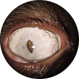
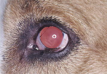
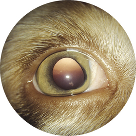
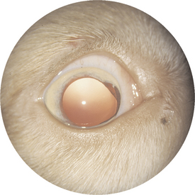
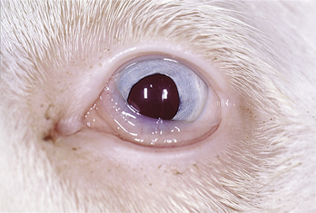
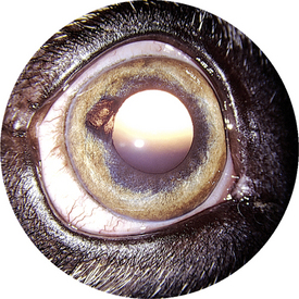
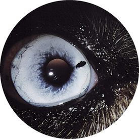
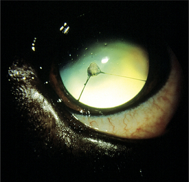
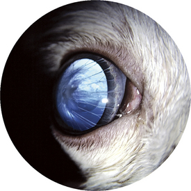
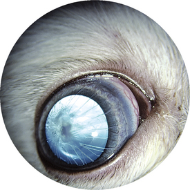
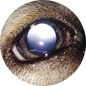
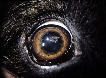
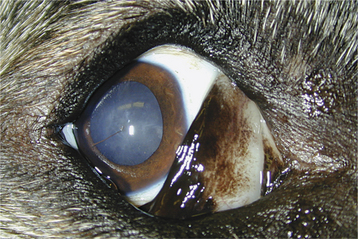
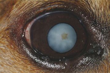
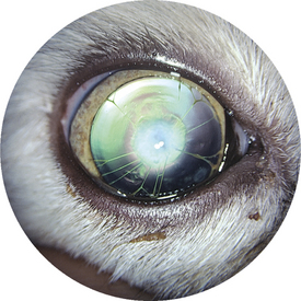
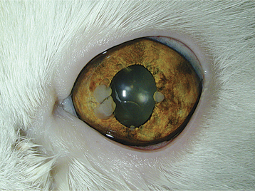
Figure 7-15 Iris-to-lens PPM with associated central cataract in a cat.
(Courtesy Dr. Robert Playter.)
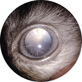
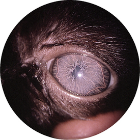
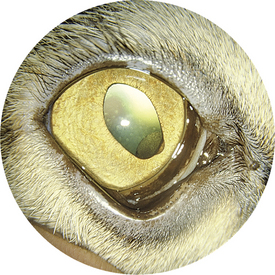
Figure 7-16 Young kitten with lipoprotein lipase deficiency and lipemia with patent PPMs and lipemic iridal vessels.
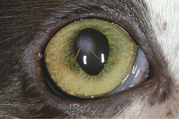
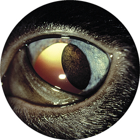
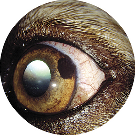
Figure 7-18 Iris-to-cornea PPM in a cat. Corneal edema may be present where the PPM touches the cornea.
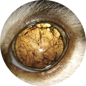
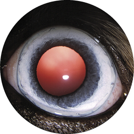
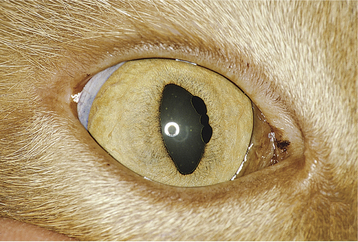
Figure 7-20 Heterochromia iridis. A marked reduction in pigmentation results in a blue color of the iris.
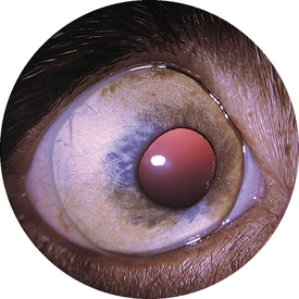
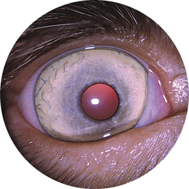
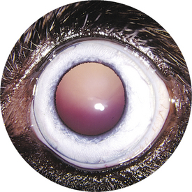
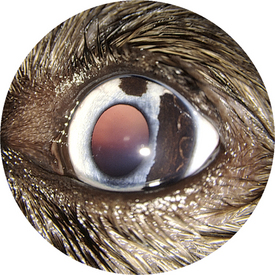
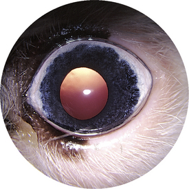
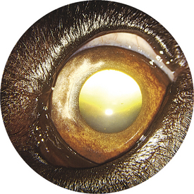
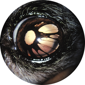
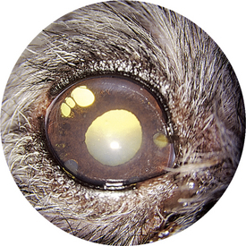
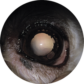
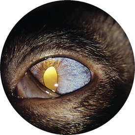
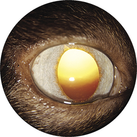
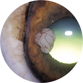
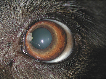
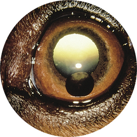
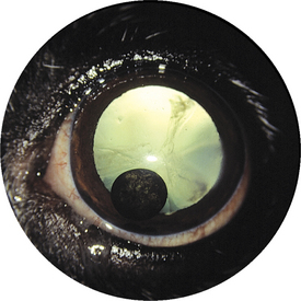
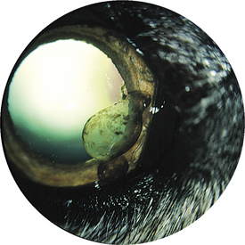
Figure 7-36 Iris cysts at the pupillary margin and in the anterior chamber in a 7-year-old Irish wolfhound.
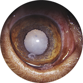
Figure 7-37 Multifocal small iris cysts at pupil margin of a dog with a cataract. Lipid keratopathy also is present.
< div class='tao-gold-member'>
Only gold members can continue reading. Log In or Register to continue
Stay updated, free articles. Join our Telegram channel

Full access? Get Clinical Tree


