Chapter 47 Uroliths and Gastroenteroliths in Camelids
Obstruction of the tubular urinary or digestive tract may cause acute colic signs and may be life threatening in any species of animal. Veterinarians are well aware of the syndromes in traditional livestock and companion animals. Similar conditions occur in zoo animals.
CONCRETIONS OF URINARY TRACT
Urinary calculi (urolithiasis; uroliths, nephroliths, bladder stones, cystoliths) are formed in either the calices of the kidney or, more often, the urinary bladder. Small uroliths may enter the ureter or urethra and cause partial or complete obstruction of urine flow.* No specific studies on the pathogenesis of urinary calculi formation in camelids have been reported. There are clinical reports of disease caused by the obstruction produced by the calculi. Zoo veterinarians must rely on information extrapolated from studies of cattle, sheep, and goats, which have urine of similar composition (Table 47-1). Box 47-1 lists the composition of urinary calculi recovered from llamas and alpacas at the Veterinary Medical Teaching Hospital, University of California. Others have reported similar findings in camels, vicu-a, and guanacos. Urethral obstruction may also occur with nonmineral concretions.
Box 47-1 Composition of Urinary Calculi*
Silicon dioxide (SiO2)—Crystobalite
Magnesium ammonium phosphate (MgNH4PO4 6H2O)—Struvite
Basic calcium phosphate (Ca5[PO4]3[OH])—Apatite
Calcium carbonate (CaCO3)—Carbonate
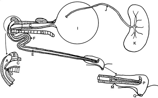
A basic understanding of the camelid urethra is required to locate sites of possible obstruction and develop approaches to management.Figure 47-1 is a diagram of the camelid urethra and associated structures. The prostate gland does not surround the pelvic urethra as in carnivores. The pelvic urethra is expansive, but at the reflection around the ischium, only a tiny orifice allows passage of urine beyond this point. The anatomy of this area is further complicated by a dorsal urethral recess that precludes any possibility of passing a catheter into the bladder from the tip of the penis. Whereas the sigmoid flexure is the probable site of the majority of bovine urethral obstructions, this is not the case in camelids. The orifice from the pelvic urethra into the penile urethra is a common site of obstruction; another site is where the penile urethra narrows as it enters the glans penis.
Clinical Signs
Uroliths in the bladder or calices of the kidney rarely cause discomfort, although large and rough-surfaced uroliths may initiate a cystitis. The signs of urethral obstruction caused by calculi vary with the stage of the disorder.9 Signs prior to bladder rupture include colic, straining stance to urinate, dribbling urine, blood-tinged urine, anuria, distended bladder, and possible pulsation of the urethra. Signs after bladder rupture are absence of colic, with depression, anorexia, anuria, uroperitoneum, and possible distention of the abdomen and uremia (muscular weakness, dehydration, dyspnea, tremors, uremic breath odor, tachycardia, recumbency, coma, death).24
Uroperitoneum may result from trauma when the bladder is distended or from rupture of the bladder after urethral obstruction. In the llama the immediate response to urine flushing into the abdominal cavity is excruciating pain. Llamas become frenzied and thrash about violently. This initial pain subsides, and the pain associated with a distended bladder disappears. Urine in the abdomen may arise from a single or multiple tears in the bladder wall, but also from seepage through the stretched-thin bladder wall. A ureter may also rupture, but such a condition has not been diagnosed in a camelid.
Diagnosis
Diagnosis is based on assessment of clinical signs, pertinent history, and special diagnostic tests. A differential diagnosis should include any disease condition in which colic is a sign. The major hematologic and serum chemistry changes found in camelids with obstructive urolithiasis include hemoconcentration, elevated blood urea nitrogen (BUN), hypophosphatemia, hypercalcemia, hypermagnesemia, hyperkalemia, hypercreatininemia, and hypochloremia.20 It is not always easy to identify the source of fluid in the abdominal cavity. Urine should have the odor of ammonia, but it may be necessary to heat the fluid to concentrate the odor before it becomes perceptible. The ability of individuals to smell ammonia varies. Abdominal fluid caused by urine has a creatinine concentration greater than 15 mg/dL (normal serum <3 mg/dL). Urine potassium is greater than185 mmol/L (normal serum <5 mmol/L). The latter is the most important biochemical urine detector.
Stay updated, free articles. Join our Telegram channel

Full access? Get Clinical Tree


