Elizabeth J. Davidson
Upper Airway Obstructions
Upper airway obstructions are among the most frequent causes of poor performance in horses, being second in frequency only to musculoskeletal injuries. Abnormalities can be static and evident at rest, or dynamic and only apparent during exercise. Complete history, signalment, physical examination, and endoscopic examination of the upper airway are necessary to ascertain the definitive cause of an upper respiratory dysfunction.
Pathophysiology of Upper Airway Obstruction
In the horse, the primary function of the upper airway is to serve as a conduit for airflow. Adequate patency of this conduit is of great importance because the horse is an obligate nasal breather, and any obstruction can negatively affect its ability to breathe. A patent upper airway is particularly essential in the exercising horse when airflow increases dramatically to meet the huge demand for oxygen by skeletal muscles (Table 20-1). These high airflows are driven by contraction of the diaphragm, which in turn creates large pressure changes in the upper airway. To remain fully open and functioning, the upper airway must counteract these large pressure fluctuations.
TABLE 20-1
Values for Respiratory Function Parameters in Horses at Rest and During Maximal Exercise
| Respiratory Parameter | Rest | Maximal Exercise |
| Respiratory rate (breaths/min) | 10-15 | 120 |
| Tidal volume (L/sec) | 3-5 | 12-15 |
| Minute ventilation (L/min) | 60 | 1400-1800 |
| Airflow (L/sec) | 3 | 75-85 |
| Peak inspiratory pressure (cm H2O) | −2 | −40 |
| Peak expiratory pressure (cm H2O) | 15 | |
| Resistance to airflow contributed by the upper airway (%) | 66 | 80 |
The rigid bony and cartilaginous structures of the upper airway are minimally affected by changes in airway dynamics. However, less rigid soft tissue and muscle structures, such as the nares, nasopharynx, and larynx, rely on neuromuscular activity to maintain stability during respiration. Any functional weakness or structural defect in these structures may result in inability to resist airway pressure gradients and airflows. This becomes increasingly important during exercise, when gradients and flows are high. Any abnormality of the upper airway that decreases the diameter or function of the airway can therefore limit respiratory function, especially in the athletic horse.
Prevalence
The true prevalence of upper respiratory obstructions is unknown because determining this would necessitate sampling of the general horse population, an undertaking that is not routinely performed. More commonly, the prevalence of obstructive disorders is reported in a population of horses presented for poor performance, exercise intolerance, abnormal respiratory noise, or a combination of these abnormalities. Racehorses have the highest requirements for airflow while training or racing and are therefore likely to have performance limitations with even modest upper airway obstructions. Exercising endoscopic studies indicate that intermittent dorsal displacement of the soft palate (DDSP) is the most common cause of upper airway obstruction in racehorses. Recurrent laryngeal neuropathy (RLN), palatal instability, pharyngeal wall collapse, and axial deviation of the aryepiglottic folds are also frequently identified. Complex dynamic upper airway obstructions are common, which highlights the importance of accurate assessment of pharyngeal and laryngeal function, not only at rest but also during exercise.
Horses engaged in less strenuous activities tolerate a greater degree of airway obstruction before performance limitation becomes apparent. In sport horses, the prevailing complaint is abnormal respiratory noise because it negatively affects the aesthetics of riding. The U.S. Equestrian Federation rulebook states that “horses must not show evidence of broken wind,” and affected horses may be penalized or disqualified from competition even if performance is not hindered. Abnormal respiratory noise or exercise intolerance can also be exacerbated by head and neck flexion, a position required or desired by certain equestrian disciplines. In show horses, pharyngeal wall collapse, palatal instability, and RLN are the most common dynamic upper airway obstructions.
Diagnosis
Accurate history and thorough physical examination are essential and should always be performed when horses are evaluated for upper respiratory tract dysfunction. Common presenting complaints include abnormal respiratory noise, exercise intolerance, and poor performance. In the racehorse, abrupt decline in performance, gradual deterioration at the end of the race, or other ill-defined forms of poor performance are also common complaints. In sport horses, the most common historical finding is that the horse makes abnormal respiratory noise. Horses competing in disciplines that require enforced poll flexion or tension may make abnormal noises while exercising in this position. Physical examination is centered on the head and neck, and should include external manual palpation of the larynx for detection of arytenoid deformities or prominence of the muscular process that develops secondary to atrophy of the cricoarytenoideus dorsalis muscle in horses with RLN. Thickening of the ventral throatlatch region or the lateral aspect of the larynx may be detected in horses that have undergone previous airway surgery.
Videoendoscopic examination of the upper airway is the gold standard for identification of upper respiratory disorders. The nasal passages, pharynx, larynx, and cranial trachea are easily examined with a flexible fiberoptic endoscope. Horses should be adequately confined for examination, and a nose twitch is usually all that is necessary to restrain the horse. Chemical sedation interferes with normal laryngeal and pharyngeal function, and its use should be avoided. Upon entry of the endoscope in the nasopharynx, the anatomy and resting function can be identified and evaluated. The position of the soft palate and ease with which it displaces dorsally should be assessed. The size and shape of the laryngeal cartilages should also be assessed. In horses with an incomplete history, the absence of a ventricle or vocal fold, or the immobility of an arytenoid cartilage, is clear indication of previous prosthetic laryngoplasty (called a tie back). Scarring of the ventral floor of the larynx and a notched caudal border of the soft palate indicates previous laryngotomy and staphylectomy. To induce arytenoid movement, the larynx is stimulated by instillation of a small volume of water through the biopsy chamber of the endoscope or by applying gentle pressure with a biopsy forceps. Immediately after the horse is induced to swallow, arytenoid cartilage function is assessed (Table 20-2). Endoscopic evaluation of the trachea from a point just caudal to the larynx to its bifurcation into the two primary bronchi (carina) completes the videoendoscopic examination.
TABLE 20-2
Grading System of Laryngeal Function
| Grade | Description |
| I | Symmetrical, synchronous arytenoid cartilage movements; full abduction and adduction can be achieved and maintained |
| II | Asynchronous arytenoid cartilage movements (flutter, hesitation); full abduction and adduction can be achieved and maintained |
| III | Asynchronous or asymmetrical arytenoid movements, or both; full abduction is not achieved and maintained |
| IV | Complete immobility of the arytenoid cartilage and vocal fold |
Complete laryngeal paralysis and structural upper airway abnormalities are easily identifiable during resting endoscopic examination. However, many obstructions are dynamic, and resting observations are notoriously unreliable in predicting exercising upper airway function. Exercising endoscopic evaluation of the upper airway is required to correctly diagnose the obstruction and is indicated in horses that do not have obvious abnormalities at rest, those with questionable laryngeal or pharyngeal function, and those that have any history of making an abnormal respiratory noise during exercise.
Specific Conditions
Alar Fold Collapse
The alar fold is a thick fold of skin extending rostrad from the ventral nasal concha. The false nostril is the space dorsal to the alar fold. During exercise, the alar fold is tensed and the space is obliterated. Inappropriate nostril dilation or redundant tissue results in alar fold collapse during exercise. The condition was originally described in Standardbred racehorses. Affected horses are normal at rest but make a loud objectionable fluttering expiratory noise during exercise. The diagnosis is made by placing a suture through the alar folds and securing them in a dorsal position. Exercising noise is greatly diminished or eliminated after suture placement. Treatment includes surgically resecting the folds or securing them during exercise. Resolution of noise and return to racing is the outcome in most surgically corrected horses.
Arytenoid Chondritis
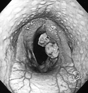
Arytenoid chondritis is an inflammatory condition of one or both arytenoid cartilages, and the etiology of this condition is unknown. Affected cartilages vary from mildly thickened to grossly deformed with mucosal ulceration, granulation tissue formation, and suppuration (Figure 20-1). In addition to chondropathy, some degree of laryngeal paralysis is frequently seen, and early in the disease process chondritis may be mistaken for RLN. Affected horses are exercise intolerant, make an abnormal respiratory noise, and often cough. During endoscopic evaluation, the opposite arytenoid should be carefully inspected because a kissing lesion (abrasion or nodule of granulation tissue) in the mucosa may result from contact with the chondritic cartilage (Figure 20-2).

< div class='tao-gold-member'>
Stay updated, free articles. Join our Telegram channel

Full access? Get Clinical Tree


