Upper Airway Disease

Chapter 16 Upper Airway Disease


CLINICAL SIGNS


The clinical signs in affected patients will vary based on species, underlying etiology, chronicity, comorbid conditions, and severity of the airway obstruction. Animals can exhibit variable degrees of dyspnea and respiratory distress. In mildly affected animals, clinical signs may include a change in voice, gagging or retching, stertor, stridor, or a dry, unproductive cough. Stertor is a low-pitched snoring sound that is generally associated with obstructive diseases affecting the nasal passages or nasopharynx. In contrast, stridor is a higher pitched respiratory noise that is generally heard during the inspiratory phase of respiration with extrathoracic obstruction and is associated with diseases affecting the larynx or trachea.1,2 The upper airway noise in these patients often increases as the severity of the obstruction progresses. Open-mouth breathing will alleviate the airway obstruction if the disease is affecting only the nasal passages or nasopharynx. Although panting is common in dogs, open-mouth breathing in cats is never normal and should alert the clinician to the presence of respiratory disease.


Hyperthermia is a common clinical sign in dogs with upper airway disease. This occurs less frequently in feline patients, likely due to their more sedentary lifestyle. Panting is one of the primary mechanisms of thermoregulation in animals; the movement of fresh air through the upper airway effectively increases heat loss through evaporation.3,4 In animals with upper airway disease, this air movement is compromised, predisposing them to hyperthermia. This hyperthermia can be extreme, and aggressive cooling may be necessary in severely affected patients (see Chapter 167, Heatstroke).5


Auscultation often reveals significant referred upper airway noise. The increased noise can be better localized by auscultating over the larynx and trachea, as well as the thorax. Inspiratory dyspnea with stertor or stridor typically results from extrathoracic obstructive diseases, and intrathoracic airway obstruction more commonly leads to expiratory dyspnea with adventitious sounds. Careful attention should be paid to thoracic auscultation to identify concurrent pulmonary parenchymal disease.


In animals with significant upper airway obstruction, severe respiratory distress and cyanosis can occur. These patients may show severe clinical signs such as an orthopneic stance when breathing, severe stridor, coughing, gasping, retching or vomiting, or collapse. In these cases, immediate anesthesia and intubation (or a tracheostomy) are imperative to minimize morbidity and mortality.


EMERGENCY STABILIZATION


Although treatment varies depending on the underlying disease process, the general emergency approach is similar in all patients with upper airway disease. As previously mentioned, these animals are fragile and can decompensate quickly, so any additional stress can be life threatening and will lead to an increase in oxygen requirements and therefore respiratory rate. This increased respiratory rate will only further exacerbate upper airway dysfunction. Early stabilization often relies on the administration of anxiolytics and sedatives (see Chapter 162, Sedation of the Critically Ill Patient). These agents should be given IM initially if the patient is not stable enough to undergo the stress of placing an IV catheter.


Acepromazine can be administered at a dosage of 0.005 to 0.05 mg/kg IV, IM, or SC. This is an excellent sedative, but it is important to remember that vasodilation and hypotension can occur, so it should be reserved for patients that are hemodynamically stable. Butorphanol is a sedative and antitussive, which can be used as a single agent or in combination with acepromazine. The dosage of butorphanol is 0.1 to 0.6 mg/kg IM, IV, or SC. In addition to sedation, patients should be placed in an oxygen-rich environment. Short-acting glucocorticoids have been recommended to reduce laryngeal and tracheal inflammation, edema, and swelling. Dexamethasone sodium phosphate at a dosage of 0.2 to 0.4 mg/kg can be administered IV, IM, or SC. In cases of suspected neoplasia, steroids should be avoided because this can complicate the diagnostic interpretation.


In patients with an elevated temperature, external cooling methods should be instituted. If sedation and oxygen therapy, with or without steroid therapy, fails to stabilize the patient, intubation is indicated. In the rare case in which intubation is not possible, an emergency tracheostomy should be performed to achieve a patent airway (see Chapter 18, Tracheostomy). When an emergency tracheostomy is not feasible, a small catheter may be passed into the trachea through the mouth or between tracheal rings and high-pressure jet ventilation or repetitive oxygen boluses from an anesthesia oxygen flow valve may be delivered until a tracheostomy is performed.


DIAGNOSTICS


Numerous disease processes can affect the upper respiratory tract. Differentiating the underlying cause in an individual patient can be challenging because the clinical signs are not pathognomonic for any single cause. Signalment, breed, history, and physical examination can often provide some information, but a detailed and thorough diagnostic approach is necessary to fully evaluate the anatomy and function of the upper airway. Even mild stress in animals with upper respiratory tract obstruction can be life threatening. In patients with mild clinical signs, less invasive testing may be possible with little or no sedation. In contrast, patients with moderate or severe airway compromise often require heavy sedation and intubation for diagnostic evaluation. In any case, the clinician should always be prepared to gain control of the airway, either via intubation or emergency tracheostomy, if necessary.


Although rarely diagnostic for a specific cause of upper airway disease, radiographs are important for evaluating both the upper and lower respiratory tracts. One ventrodorsal and two lateral views of the thorax should be taken to evaluate for metastatic disease, aspiration pneumonia, or noncardiogenic pulmonary edema. Thoracic radiographs are also used to evaluate the trachea and look for intrathoracic masses. Cervical radiographs may show evidence of laryngeal or pharyngeal neoplasia (Figure 16-1), radiodense foreign bodies, or extramural masses causing compression of the larynx or trachea. In patients with nasal or nasopharyngeal disease, skull radiographs (or computed tomography) may be indicated to evaluate the nasopharynx, the bullae, and the external canal and petrous temple bone. Unlike thoracic and cervical radiographs, patients undergoing skull radiography require general anesthesia for proper radiographic positioning.
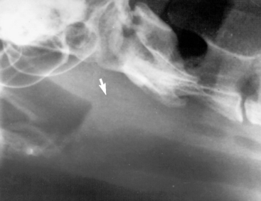


Laryngeal examination is an essential part of the diagnostic evaluation in any patient with clinical signs attributable to upper airway disease. The laryngeal examination is used to evaluate the function and movement of the rima glottidis, including the vocal cords and arytenoid cartilages. The larynx is visualized to assess for thickening or irregularities, and the soft palate and laryngeal saccules should be identified and evaluated.


After evaluation of the upper respiratory tract, an endotracheal wash should be considered, particularly in any patient with signs of lower airway disease. The patient should be intubated with a sterile endotracheal tube and sterile saline aliquots (3 to 5 ml for cats; 5 to 7 ml for dogs) are injected into the endotracheal tube through a sterile red rubber catheter. The patient is given gentle coupage and the fluid suctioned via the red rubber catheter or a sterile suction catheter. The fluid obtained should be submitted to a diagnostic laboratory for fluid analysis and bacterial culture and sensitivity. Additional cultures may be indicated based on cytologic findings.


Endoscopy is required for evaluation of the upper airway caudal to the epiglottis. Endoscopy is used to identify nasopharyngeal stenosis, nasopharyngeal abscesses or masses, nasopharyngeal polyps, or extramural compression. The endoscope can be retroflexed to evaluate the caudal nasal cavity. Finally, tracheoscopy is helpful in evaluating for tracheal collapse, tracheal tumors, extramural compression, or tracheal foreign bodies.


In cases with an irregularity, thickening, or mass effect associated with the larynx or pharynx identified on laryngeal examination, biopsy is always required for a definitive diagnosis because neoplasia and inflammatory disease are indistinguishable on gross examination.


Computed tomography of the skull can be an invaluable diagnostic test for nasal and nasopharyngeal disease. Although it cannot be used for functional abnormalities such as laryngeal paralysis, it is an excellent tool for evaluating the nasal and nasopharyngeal area for tumors or osteolysis, polyps, abscesses, stenosis, or collapse. As with skull radiographs, general anesthesia is required for adequate positioning and to avoid movement artifacts.

Stay updated, free articles. Join our Telegram channel

Sep 10, 2016 | Posted by in SMALL ANIMAL | Comments Off on Upper Airway Disease

Full access? Get Clinical Tree

Get Clinical Tree app for offline access