32 Uncomplicated crown fracture with periapical complications
ORAL EXAMINATION – CONSCIOUS
The dog was aggressive when the head was approached and conscious oral examination was not possible.
ORAL EXAMINATION – UNDER GENERAL ANAESTHETIC
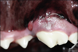
In summary, examination under general anaesthesia identified the following:
RADIOGRAPHIC FINDINGS
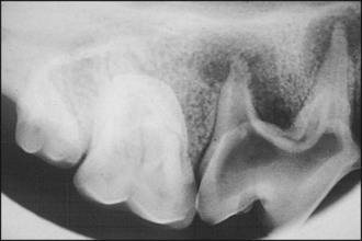
The diameter of the pulp system of 108 was wide and the apices of the roots were open, indicating that the injury occurred during tooth development in the first year of life. There was also periapical inflammation with bone destruction and external root resorption (see Fig. 32.2 for details of the radiographic findings for 108).
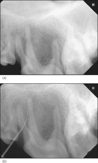
The diameter of the pulp system of 208 was narrow and the apices were closed, i.e. it was a mature tooth, as expected in a 9-year-old dog. There was periapical inflammation with bone destruction. The drainage tract was shown to originate from the mesiopalatal root (see Fig. 32.3a, b for details of the radiographic findings for 208).

Figure 32.3 Radiographs centring on 208.
Stay updated, free articles. Join our Telegram channel

Full access? Get Clinical Tree


