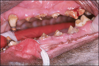
38 Enamel dysplasia
ORAL EXAMINATION – CONSCIOUS
The dog would not allow access to her face at all, so conscious oral examination was not performed.
ORAL EXAMINATION – UNDER GENERAL ANAESTHETIC
In summary, examination under general anaesthesia identified the following:
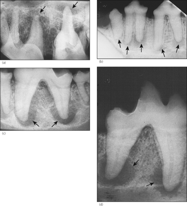
RADIOGRAPHIC FINDINGS
All teeth, except incisors and canines, were affected by pulp and periapical pathology (see Fig. 38.2).
THEORY REFRESHER
Enamel dysplasia (hypoplasia) may be defined as an incomplete or defective formation of the organic enamel matrix of teeth. The result is defective (soft, porous) enamel. It can be caused by local, systemic or hereditary factors. Depending on the cause, the condition can affect one or only a few teeth (localized form), or all teeth in the dentition (generalized form). It is essential to remember that enamel dysplasia results only if the defect occurs during the formative stage of enamel development, i.e. during amelogenesis. Thus, the defect occurs before the tooth erupts into the oral cavity. Crown formation lasts from the 42nd day of gestation through to the 15th day postpartum for the primary teeth, and from the second week through to the third month postpartum for the permanent teeth of dogs and cats. Depending on the time of the insult, enamel dysplasia will affect primary and/or permanent teeth. Only those areas of enamel undergoing active formation during the period of the insult will be affected. This is seen clinically as bands of dysplastic enamel encircling the crown, with areas of normal enamel elsewhere on the tooth.


