Ultrasonographic Examination of the Upper Airway

CHAPTER 52 Ultrasonographic Examination of the Upper Airway


Clinical evaluation of the upper airway of horses with the presenting complaint of poor performance or abnormal upper respiratory noise is typically initiated with a thorough physical examination to localize the problem. This includes assessment for enlargement or obstruction of the nasal passages or other evidence of sinonasal disease, including nasal discharge and physical deformation, and palpation for evidence of acquired or congenital diseases. Acquired diseases include tracheal deformation, neurogenic atrophy of the dorsal cricoarytenoideus muscle, and thrombophlebitis, whereas congenital disease is exemplified by fourth branchial arch defect. Physical examination may be normal or, in many instances, might not fully characterize the problem, necessitating further diagnostic tests.


Of these, endoscopy of the horse at rest is the most commonly used diagnostic method. It has the advantages of being widely available and relatively noninvasive. Further, it offers both structural and functional information about the upper airways. However, evaluation is limited to assessment of luminal structures, and functional information gained is not always representative of airway performance at exercise. Video endoscopy during exercise is the best means of evaluating the upper airway for dynamic and multiple obstructions and may reveal lesions that are not apparent in a resting examination. However, there are several limitations to using video endoscopy at exercise, including the cost of treadmill examination, limited availability of treadmill facilities, and a view that is limited to luminal structures, with palpation being the only means of assessing nonluminal structures. Laryngeal ultrasound was introduced as an added diagnostic technique for the equine upper airways to address some of these limitations.


Ultrasound examination of the larynx offers several advantages: it can be performed on the standing horse, yields both structural and functional information, and allows the nonluminal aspect of the upper airway to be evaluated. Specifically one can assess the following structures: portions of the hyoid apparatus, laryngohyoid position, strap muscles of the neck, laryngeal and perilaryngeal musculature, and laryngeal cartilages (except the epiglottis). Because the upper airway anatomy is complex, a thorough understanding of ultrasonographic imaging principles and equine laryngeal anatomy is necessary to develop competency in this technique.


This chapter provides a practical introduction to the technique and its clinical applications. At the time of publication, research is ongoing concerning use of ultrasound for diagnosing important upper airway conditions.


EQUIPMENT AND PREPARATION


Although there is a wide variety of ultrasound equipment available to the equine practitioner, much of our experience has been gained using a Phillips HDI 5000 or 3000 (Philips Medical Systems, Bothell WA 98041). Ideally the examination is performed with high-resolution transducers, and two different transducers are generally needed to enable imaging of all structures and facilitate accurate measurement. One transducer should have a flat footprint for accurate percutaneous measurements, such as a linear-array transducer typically used to image tendons. However, these types of transducers are often bulky, with a footprint so large that the caudal edge of the mandible blocks an image of the lateral laryngeal structures, and a second smaller convex transducer is often necessary. We typically use a 12.5-MHz linear array transducer (38 mm) and a 8.5-MHz convex (14-mm) transducer during a systematic laryngeal examination. The highest-frequency probe that is capable of penetrating the desired tissue depth should be used for each portion of the examination to maximize image resolution. In some instances, additional lower-frequency probes may be required to obtain an image of deeper structures or penetrate swollen tissues. Although sector scanner probes are ideal for imaging through narrow acoustic windows, their close-up image display has a limited width, which impedes imaging of some superficial structures, and their use is generally not preferred.


It is preferable to examine the horse while restrained in stocks and wearing a standard halter, so that the handler can limit its head movements. The examination is performed with the horse unsedated to allow accurate evaluation of arytenoid cartilage and vocal-fold movement and of the laryngohyoid position. Hair clipping is generally not required except for heavily coated draft breeds. Alcohol is the preferred acoustic coupling agent; however, acoustic coupling gel may be used instead. In some instances, skin-associated artifacts that reduce image quality and can impede image interpretation have been encountered. These seem to occur when the examination takes longer and the alcohol begins to dry on the hair (alcohol drying effect) or the horses undergo strenuous exercise before ultrasound. The sweat generated during exercise or the cold hosing following treadmill testing may be responsible for reducing image quality.


ULTRASOUND TECHNIQUE


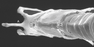
As with any ultrasound examination, it is important to evaluate the larynx in a systematic manner. This is facilitated by a standardized examination that uses specified imaging locations termed acoustic windows. This is particularly important in the laryngeal region because gas, bone, and mineralized cartilage all create acoustic shadows that prevent imaging. The use of standard acoustic windows allows the ultrasonographer to avoid bone and gas impedance, develop familiarity with the normal appearance at each site, clearly communicate abnormal findings by location, and ensure that all relevant structures are imaged each time. It is recommended that the ultrasonographer proceed sequentially through the five acoustic windows described and label any stored images appropriately (Figure 52-1). Beginning on the ventral aspect of the throat and proceeding from rostral to caudal, the following acoustic windows are evaluated: rostroventral (rostral to the basihyoid bone), midventral (region between the basihyoid bone and thyroid cartilage), and caudoventral (imaged through the cricothyroid membrane and extending caudally to the first few tracheal rings). On completing the examination through the ventral windows, imaging proceeds with the right and left caudolateral windows just caudal to the angle of the mandible at the lateral aspect of the larynx on each side.


Ultrasonography is usually performed in both longitudinal and transverse planes for the ventral windows and in a rostrocaudal plane for the lateral windows. The routine examination beyond the laryngeal region may need to be modified for certain lesions to include the root of the tongue, mandibular or retropharyngeal lymph nodes, salivary or thyroid glands, or jugular veins. Although dorsal cricoarytenoid muscle assessment has much relevance to laryngeal disease with regard to laryngeal hemiparesis and hemiplegia, a reliable technique for evaluating this muscle has not been developed.


May 28, 2016 | Posted by in EQUINE MEDICINE | Comments Off on Ultrasonographic Examination of the Upper Airway

Full access? Get Clinical Tree

Get Clinical Tree app for offline access