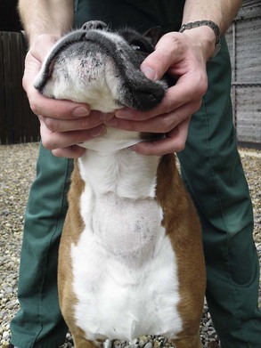
Thyroid Tumors

Chapter 89


Thyroid Tumors


The thyroid gland is a bilobed structure in cats and dogs, sometimes with an isthmus connecting the two lobes; it spans a region from the fifth to the eighth tracheal ring, and the right lobe usually is more cranial than the left. In normal animals, the lobes are lateral to and closely associated with the trachea. The gland is extremely well vascularized in both cats and dogs, especially when neoplastic.


Ectopic thyroid tissue is common and can be found from the base of the tongue to the heart base, anywhere vestigial tissue may be present from embryologic development. Neoplasia can arise in this ectopic tissue.


The thyroid gland is composed mostly of two cell types, both of which can become neoplastic. Thyroid epithelial cells—the cells responsible for synthesis of the thyroid hormones—are arranged in thyroid follicles around a central space filled with colloid. In spaces between thyroid follicles are found the parafollicular or C cells, which secrete the hormone calcitonin.


Palpable masses can be caused by hyperplasia, adenoma, or adenocarcinoma of the thyroid gland. In cats, benign adenomatous hyperplasia is a common presentation in older animals, with the functional tissue giving rise to the signs associated with hyperthyroidism. In contrast, the vast majority of thyroid tumors in dogs are nonfunctional and malignant, with benign lesions usually being an incidental finding at postmortem.


In humans, thyroid tumors can be seen as part of two multiple endocrine syndromes referred to as multiple endocrine neoplasia type 1 and type 2 (MEN1, MEN2). It is not clear that cats or dogs have shown evidence of a truly analogous syndrome; however, it is important to note that thyroid tumors have been seen in dogs that had developed tumors of other histologic types, and so it is especially important to examine the animal thoroughly at presentation for other masses. The additional distinct tumors were not in other endocrine organs.


Canine Thyroid Carcinoma


Thyroid cancer represents between 1% and 4% of all canine tumors. Tumors arising from the cells lining the follicles are known as follicular carcinomas, which are further subclassified as papillary, compact (solid), or anaplastic, whereas those arising from the parafollicular cells are called medullary thyroid carcinomas or C-cell carcinomas and are less common.


Affected dogs usually are 9 to 10 years or older. Boxers, beagles, golden retrievers, and Siberian huskies have been found to be more commonly affected in some studies. A mixed-breed family of dogs (three out of four littermates) with a significant pedigreed Alaskan malamute component in their breeding developed medullary thyroid carcinomas (Lee et al, 2006). There is no reported sex predisposition.


Presentation


Clinical signs depend on where the tumor is located and whether the mass is functional. The vast majority of the lesions are nonfunctional and are situated in the area of the thyroid gland; therefore a noticeable mass is by far the most common presenting sign (Figure 89-1). The average time to diagnosis is 1 to 2 months from owner appreciation of the mass. It is important to assess mobility of the mass. Roughly one third to one half of thyroid carcinomas are freely moveable at presentation, and freely mobile thyroid carcinomas have different treatment options and different prognoses from fixed carcinomas.


Other clinical signs frequently reported are associated with the primary mass and include dysphonia, dysphagia, and cough. Dyspnea can be associated with either the primary mass, which may press on the trachea, or with lung metastases. Sometimes signs can be present that are consistent with laryngeal paralysis because the mass can damage the recurrent laryngeal nerves. Rarely, facial edema can be seen in association with impaired venous return.


Most dogs with thyroid carcinomas are euthyroid; however, thyroid function is affected in a small percentage of cases. Therefore a dog with clinical signs consistent with hyperthyroidism (polyuria, polydipsia, weight loss, increased appetite, muscle atrophy, hypertension, nervousness) should undergo routine thyroid function testing. Additionally, hypothyroidism may be diagnosed in some, which may precede the tumor’s development or be caused by destruction of normal thyroid tissue. Unfortunately, there are few studies that report thyroid hormone assay results in dogs with thyroid carcinoma so it is difficult to say with any accuracy what percentage of dogs are hypothyroid, euthyroid, or hyperthyroid at presentation.


Medullary carcinomas usually are nonfunctional, but calcitonin-producing tumors causing hypocalcemia have been reported.


Diagnosis


It has been suggested that a palpable mass in the thyroid of a dog has a 85% to 90% probability of being malignant. Differential diagnoses include abscess, granuloma, foreign body, lymphadenitis, lymph node metastases, lymphoma, and subcutaneous tumors such as soft-tissue sarcoma and mast cell tumor.


Thyroid tumors are extremely vascular, and thus the first approach to diagnosing such a mass may be fine-needle aspiration, without suction, followed by digital pressure after the needle is withdrawn. Because these lesions are well vascularized it is not surprising that blood contamination is a problem. Ultrasonographic guidance may be used to minimize the risk of sampling the most well-vascularized areas.


Surgical biopsy can be challenging due to the high risk of hemorrhage, and so a more pragmatic approach is to assess the mass for surgical resectability and obtain a diagnosis at the time of surgery. This assessment may involve palpation, ultrasonography, or computed tomography or magnetic resonance imaging to be sure that the mass is not attached to other structures.


Once histopathologic evaluation can be undertaken, any diagnostic dilemma as to tumor type can be resolved by immunohistochemical analysis. This may be especially useful if ectopic tissue is involved. Thyroid follicular tumors stain positive for thyroglobulin. Medullary tumors can be confused with compact follicular tumors, and an immunostain for calcitonin can be used to differentiate the two, with medullary tumors staining positive for calcitonin.

< div class='tao-gold-member'>

Stay updated, free articles. Join our Telegram channel

Jul 18, 2016 | Posted by in PHARMACOLOGY, TOXICOLOGY & THERAPEUTICS | Comments Off on Thyroid Tumors

Full access? Get Clinical Tree

Get Clinical Tree app for offline access