Chapter 8 Third Eyelid
ANATOMY AND PHYSIOLOGY
The third eyelid (or nictitating membrane) is a mobile, protective, and glandular structure lying between the cornea and the lower eyelid in the medial portion of the inferior conjunctival sac (Figure 8-1).
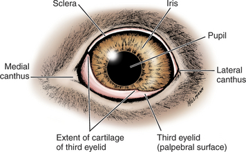
Figure 8-1 Diagram of the eye showing normal position of the third eyelid.
(Modified from Evans HE [1993]: Miller’s Anatomy of the Dog, 3rd ed. Saunders, Philadelphia.)
The third eyelid (Figure 8-2) consists of the following:
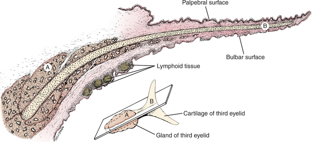
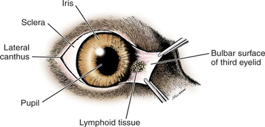
The T cartilage provides essential rigidity to the third eyelid. Its “horizontal” arm lies parallel to and about 1.5 mm from the leading edge of the third eyelid. The “vertical” arm runs perpendicular to the free edge and at its base is encircled by the gland of the third eyelid (see Figure 8-2). The gland of the third eyelid is seromucoid and produces up to 50% of the normal tear film in dogs. In the dog, this gland has both adrenergic and cholinergic innervation, with cholinergic being the denser. In the pig and many rodents a portion of the gland of the third eyelid or a separate gland (the Harderian gland) is found deeper within the orbit. The cartilage and gland are covered on both bulbar and palpebral surfaces by conjunctiva that is tightly adherent at the free margin of the third eyelid but looser over the base and gland. The free margin and a portion of the anterior face of the third eyelid are often but not always pigmented. Lymphoid follicles, which are pinkish red, are normally present beneath the conjunctiva on the bulbar surface of the third eyelid (Figure 8-3).
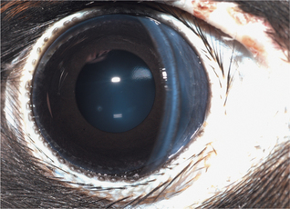
In birds the third eyelid is almost transparent and is under voluntary control (Figure 8-4). It sweeps over the globe in a ventromedial direction from the dorsolateral quadrant, although there is some species variation in direction of movement. The third eyelid in birds does not have a gland associated with it.
The third eyelid has the following important functions:
Therefore removal of the third eyelid or its gland predisposes to the following problems:
EXAMINATION
The clinician can easily examine the palpebral surface of the third eyelid by digitally retropulsing the globe through the upper lid. The bulbar surface is examined after application of topical anesthesia and the use of forceps or mosquito hemostats to grasp the leading edge of the third eyelid just outside the horizontal arm of the cartilage. The membrane can then be reflected to examine the bulbar surface and the space between the third eyelid and the globe (see Figure 8-3). This is a common site for foreign bodies to become lodged. The bulbar surface is normally follicular and may become more so with so-called follicular conjunctivitis (see Chapter 7).
DISEASES OF THE THIRD EYELID
Because the third eyelid has two surfaces of conjunctiva and is intimately associated and confluent with the rest of the conjunctiva, it is predictably involved in many conjunctival disorders. These are discussed more fully in Chapter 7. This chapter emphasizes conditions peculiar to the third eyelid.
Stay updated, free articles. Join our Telegram channel

Full access? Get Clinical Tree


