Chapter 120The Western Performance Horse
 The Cutting Horse
Jerry B. Black and Robin M. Dabareiner
Diagnosis and Management of Specific Lameness
Selected Lameness of the Tarsus
Selected Lameness of the Stifle
 The Roping Horse
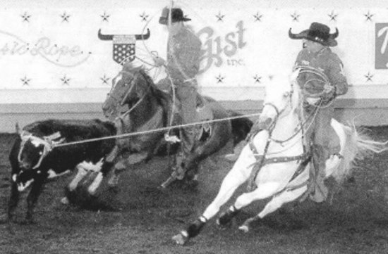
Team Roping Horse
Historical Data and Decreased Performance
It is imperative for a clinician to know whether a team roping horse is used primarily as a heading or heeling horse. In addition, a thorough description of the owner’s complaint of a change in or decreased performance provides valuable insight into the underlying problem, bearing in mind that often in the early stages little or no lameness may be present. Eighty-nine of 118 (75%) team roping horses had a history of lameness, and 29 (25%) had an owner complaint of decreased or altered performance.1 The type of performance change differed between horses used for heading or heeling (Figures 120-1 and 120-2).