Shock Wave Therapy

Chapter 96Shock Wave Therapy


Historical Perspective


Early studies in people in which shock waves (SWs) were used to disintegrate ureteral stones resulted in radiologically evident remodeling of the pelvis.1 These findings sparked the initial studies investigating the use of SWs in orthopedic applications. These studies and anecdotal clinical use resulted in the initial five standard indications used in human medicine: (1) calcifying tendonitis of the shoulder (tendinosis calcarea), (2) tennis elbow (lateral epicondylitis), (3) golfer’s elbow (medial epicondylitis), (4) heel spurs (plantar fasciitis), and (5) pseudarthrosis. Focused shock wave therapy in horses started in Germany in 1996, and applications were primarily based on those from human medicine.2 Because of positive experiences treating people with insertional desmopathies, the first equine application was the use of shock wave therapy (SWT) in horses with proximal suspensory desmitis. Initial clinical responses were positive; therefore SWT was attempted in numerous other equine conditions, including navicular syndrome and distal hock joint pain.3,4 Initially, the probe was positioned behind the navicular bone from the heel, but when ultrasonographic imaging showed that the distal sesamoidean impar ligament could be seen through the frog, this location was then used to administer SWT to the navicular bone and associated structures.


The first equipment was large, requiring water cycling and degassing, and use was limited to horses under general anesthesia. The development of more affordable, portable, and durable equipment led to an initial expansion of its use and applications. However, as further knowledge was gained in equine medicine and surgery, the use of SWT has contracted somewhat, and attention is now paid to applications that consistently provide good clinical outcomes.


Shock Wave Generators


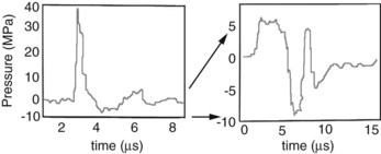
Two distinctly different pressure waves have been loosely described as SWT, SWs and radial pressure waves (RPWs). True SWs are pressure waves that meet specific physical parameters, including a rapid rise time (within nanoseconds), high peak pressures, and a gradual decrease in pressure over a few milliseconds, often with a negative pressure component (Figure 96-1 and Table 96-1).5 SWs occur naturally associated with lightning, planes breaking the sound barrier, or explosions. Electricity is used as a driving energy source to generate SWs used for medical purposes. RPWs are generated by pneumatically powered sources that drive a metal rod to strike a plate in contact with the skin surface. RPWs have slower rise times and lower peak pressures than SWs. Differences between SWs and RPWs are important because they may not affect tissue similarly; consequently, the type and equipment used are critical when evaluating the effect on tissues and efficacy of treatment. Editors’ note: However, published results in the horse have to date shown little difference in efficacy. A comprehensive review of SWs and RPWs, along with a description of equipment currently available, was recently published.6


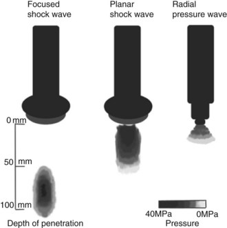
TABLE 96-1 Comparison of Shock Waves and Radial Pressure Waves


SHOCK WAVES RADIAL PRESSURE WAVES
image
Rise time 5-10 ns 50 µs
Focusing Yes No*
Maximum pressure location At focal point On surface
Energy loss Minimal through fluid/tissue Loss proportional to the square of the distance
Peak pressure ≈100 MPa ≈10 MPa
Energy flux density 0-3 mJ/mm2 0-0.3 mJ/mm2

* Includes “focused” end piece.


Reprinted with permission from Robinson EN, Sprayberry KA: Current therapy in equine medicine, ed 6, St. Louis, 2009, Saunders.


Dose Dependence


There is a dose effect of SWT that is a combination of the energy flux density (EFD) and the number of pulses. The EFD (mJ/mm2) is the amount of energy (millijoules) in the focal area (mm2). There is a lower limit of EFD that must be reached for SWT to be effective and an upper level over which EFD is high enough to create damage at any number of pulses. This range depends on species, tissue being treated, waveform, and generator.


It is difficult to transpose doses from other species to horses. When 1000 pulses at an EFD of 0.28 mJ/mm2 was applied to rabbit Achilles tendons, there was a useful inflammatory reaction; but there was inflammation and tendon necrosis at 0.6 mJ/mm2.7 However, EFDs of 0.6 mJ/mm2 or more are used in horses without complications. The minimal dose to achieve the desired effect is not known, and we may not need to use such high levels. There is a current trend to use low EFDs for a number of applications. One thousand pulses at 0.18 mJ/mm2 stimulated neovascularization of the tendon-bone junction in dogs.8 A dose of 200 pulses at 0.12 mJ/mm2 was better than 500 pulses in an Achilles tendonitis model in rats.9 At this time, the ideal energy levels and pulse numbers remain empirical in most equine applications.


The frequency (pulses/sec) of the pulse application appears unimportant, although SWT using lower frequencies takes longer. From a biological standpoint, frequency of all equine SWT applications is relatively slow, and tissue heating or other untoward biological effects seen at high rates do not occur, although white hair occasionally develops at treatment sites.


Applications


Bone


Early studies of SWs on bones yielded variable results and were confusing. Very-high-energy treatment of rodent and rabbit bones resulted in physical disruption of the bone and periosteum, which led to the thought that bone remodeling in response to SWT was the result of physical damage.10,11 In other studies using more appropriate tissue and in different species, bone remodeling occurred without physical disruption. The ability for SWT to stimulate bone healing resulted in an interest to use SWs for the management of nonunion fractures. In one study, nonunion fractures were created in 10 dogs using segmental radial osteotomies.12 Five dogs were treated with 4000 SWs at 0.54 mJ/mm2, and five served as untreated controls. All treated dogs had osseous union by 12 weeks compared with only one untreated control dog.12 The first widespread musculoskeletal use of SWs in people was for management of nonunion fractures. Clinical trials have consistently demonstrated bone healing following SWT in delayed union fractures and nonunion fractures. Wang reported 80% success13; Schaden, 75% success14; and Rompe, 72% success—results that were remarkably consistent.15


The mechanisms of SW-induced osteogenic stimulation are currently being investigated. There were increased osteoprogenitor colony-forming units from the marrow of rat femora 24 hours after treatment with 500 SWs at 0.16 mJ/mm2.16 A dose response was seen with the highest supernatant concentrations of transforming growth factor–β1 (TGF-β1) after 500 SWs; lower numbers of pulses were ineffective, and higher numbers were inhibitory. The same treatment protocol (500 SWs at 0.16 mJ/mm2) was administered to rats with segmental bone defects and resulted in increased expression of TGF-β1 and vascular endothelial growth factor–A (VEGF-A) mRNA and increased osteoprogenitor cells.17 In both studies, it appeared that SWT stimulated osteogenesis without physical disruption of the bone. Subsequent studies have shown that SWT increases concentrations of VEGF and bone morphogenic protein–2, as well as neovascularization and ultimately an increase in load to failure in a rabbit femoral fracture model.18


The application of SWT to bone was the initial musculoskeletal application in people and horses. The applications and outcomes in human medicine have grown and are positive. The applications of SWs in horses have not been pursued as aggressively as may have been expected. One initial concern was the potential for the creation of microfractures that could weaken the bone after treatment, a theory that has some support. The application of 9000 pulses of RPWs at 0.175 mJ/mm2 or 9000 SWs at 0.15 mJ/mm2

Stay updated, free articles. Join our Telegram channel

Jun 4, 2016 | Posted by in EQUINE MEDICINE | Comments Off on Shock Wave Therapy

Full access? Get Clinical Tree

Get Clinical Tree app for offline access