The Carpal Canal and Carpal Synovial Sheath

Chapter 75The Carpal Canal and Carpal Synovial Sheath


Anatomy


The carpal canal encloses the carpal synovial sheath, which contains the superficial (SDFT) and deep (DDFT) digital flexor tendons. The dorsal wall of the carpal canal is formed by the common palmar ligament of the carpus, which is a thickened part of the fibrous joint capsule that extends distally as the accessory ligament of the DDFT (ALDDFT). Proximally the accessory ligament of the SDFT (ALSDFT) forms the medial wall of the canal. Laterally the carpal canal is formed by the accessory carpal bone and the accessorioquartale and accessoriometacarpeum ligaments extending distally. The caudal antebrachial fascia, flexor retinaculum, and palmar metacarpal fascia form the palmar aspect of the canal.


The carpal synovial sheath extends from 7 to 10 cm proximal to the antebrachiocarpal joint to the midmetacarpal region. The proximal recess is wide and extends between the ulnaris lateralis and lateral digital extensor muscles laterally, but it is firmly supported on the medial aspect by the antebrachial fascia. The distal recess extends between the DDFT and its accessory ligament. If the carpal sheath is distended, swelling can be seen on the lateral aspect of the distal antebrachium and between the DDFT and its accessory ligament, medially or laterally in the metacarpal region.


The ALSDFT arises from the caudomedial aspect of the radius about 10 cm proximal to the antebrachiocarpal joint. The ALSDFT is a fibrous fan-shaped band that merges with the SDFT at the level of the antebrachiocarpal joint and prevents overload of the SDFT muscle during overextension of the metacarpophalangeal joint. After desmotomy of the ALSDFT in cadaver specimens, strain on the SDFT is increased.1 At the level of the distal aspect of the radius is an extension from the lateral aspect of the sheath wall between the SDFT and DDFT. At the level of the accessory carpal bone is a mesotendon extending from the lateral aspect of the DDFT to the sheath wall. In clinically normal horses, the amount of fluid within the carpal sheath varies, but it is usually the same bilaterally in each horse.


Fluid within the sheath may be seen readily by ultrasonography between the DDFT and its accessory ligament in normal horses, with no palpable distention of the sheath wall.2 Within the proximal part of the carpal sheath, the SDFT and DDFT contain muscular tissue and therefore have hypoechoic regions within them on ultrasonographic examination.3-5 However, the ALSDFT is uniform in its echogenicity.4,5 The position of the accessory carpal bone prohibits ultrasonographic examination from the caudal aspect of the carpus. The carpal sheath and its contents are evaluated most easily from the distal caudomedial aspect of the antebrachium and carpus and the palmar aspect of the proximal metacarpal region. The heterogeneous echogenicity of the digital flexor tendons proximally can make definitive diagnosis of a tendon lesion difficult, but comparison with the contralateral limb may be helpful. Endoscopic evaluation may yield further information and permit surgical debridement of torn fibers (see Chapter 24).


The transverse ridge of the distal aspect of the radius is at about the same level as the distal physis. Irregular roughening of this ridge may be seen radiologically in normal horses and should not be confused with entheseous new bone associated with tearing of the attachment of the ALSDFT further proximally.


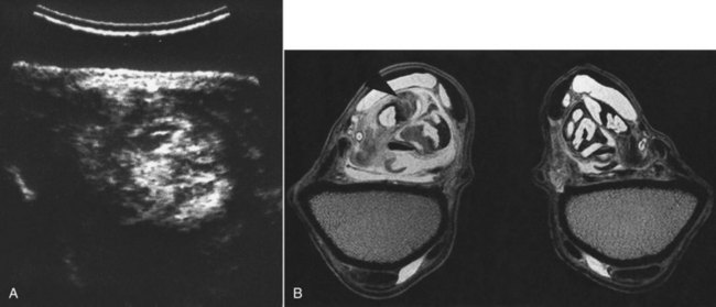
Occasionally abnormalities cannot be detected using conventional imaging techniques and magnetic resonance imaging (MRI) is required (see Figure 75-1, B). Normal MRI anatomy of the carpal region has recently been described.6


Clinical Signs


Lameness associated with the carpal synovial sheath is usually accompanied with some distention of the sheath. There may be generalized thickening in the region of the flexor retinaculum. The horse may have restricted flexibility of the carpus, with pain on passive flexion. Alternatively, the horse may resent full extension of the carpus. Rarely, increased pressure within the carpal sheath may result in compromised blood flow within the median artery and reduction in arterial pulse amplitudes in the more distal part of the limb.7 Palpation of the structures within the proximal part of the carpal sheath is not possible, but the SDFT, DDFT, and ALDDFT should be assessed carefully in the metacarpal region. Lameness varies from mild to severe and usually is improved by intrathecal analgesia. Clinical investigation should include radiographic and ultrasonographic examinations. In the absence of effusion of the carpal sheath, intrathecal analgesia should be performed to verify the source of pain and the clinical significance of any postulated lesion.

Jun 4, 2016 | Posted by in EQUINE MEDICINE | Comments Off on The Carpal Canal and Carpal Synovial Sheath

Full access? Get Clinical Tree

Get Clinical Tree app for offline access