Arthroscopic Examination

Chapter 23 Arthroscopic Examination


Arthroscopic surgery is arguably the most important advance in management and one of the most important in diagnosis of equine joint disease. Arthroscopic surgery has been a mainstay in managing joint disease since the early 1980s and has mostly replaced arthrotomy. An effective equine surgeon cannot lack extensive arthroscopic surgical experience, and lameness diagnosticians must understand indications and limitations of the technique. Innovators have used the same instruments for bursoscopy and tenoscopy (see Chapter 24). For a complete description of procedures, instrumentation, and principles of arthroscopy, the reader is referred to Diagnostic and Surgical Arthroscopy in the Horse.1


Advantages and Disadvantages of Arthroscopic Surgery Compared with Arthrotomy


Arthroscopic surgery offers several advantages compared with arthrotomy; however, this type of surgery also has some disadvantages. Both are discussed in the following text.


Advantages


Reduced Trauma and Morbidity


Arthroscopic examination is less traumatic and causes less morbidity. Arthroscopic examination allows surgery to be performed through small incisions and requires less surgical exposure and damage to overlying soft tissues, upholding a time-honored principle of limiting trauma. Little pain is observed in horses after arthroscopic surgery compared with arthrotomy, and many horses appear to ambulate normally. However, in an unblinded study of horses after arthroscopy under general anesthesia, horses showed mild but significant increases in discomfort compared with pain-free controls.2 Complications such as wound dehiscence and seroma formation are minimal. Horses often show pronounced lameness for several days after arthrotomy. After arthroscopic examination and surgery, owners and trainers expect horses to return to full work soon after surgery, an idea that is fueled by widespread reports of human athletes returning to professional sports quickly after arthroscopic surgery. A general misunderstanding is that incision size is the limiting factor. Although arthrotomy does cause short-lived lameness after surgery, the fear that it will delay onset of training is unfounded because the underlying lesion dictates recovery time. Although not recommended, horses with mild conditions such as osteochondritis dissecans, effusion but no lameness, and those that receive prophylactic arthroscopic surgery can resume training a few weeks after arthroscopic examination or arthroscopic surgery. Arthroscopic surgery can be performed within weeks before a sale, and if hair was not clipped, surgical sites are barely noticeable.


Disadvantages


Improper Case Selection


Case selection is the most important disadvantage. Poor case selection can make arthroscopic surgery difficult and disappointing. Although much information can be gained by arthroscopic examination of any joint, prognosis should be carefully considered when operating on joints in which extensive osteoarthritis exists. Inadequate communication before surgery may leave owners and trainers with too high expectations of results. Often they have little appreciation for the magnitude of cartilage damage and osteoarthritis (OA), and they only see or hear about the chip fracture(s) visible radiologically. Owners and trainers tend to want to do something, but removal of osteochondral fragments from a carpus or fetlock joint with hopeless OA can be time consuming, cause instrument failures, and, worse, often results in a poor outcome. Poor prognosis associated with some conditions must be well communicated to owners before surgery. For example, we recently reviewed the results of surgical and conservative management of scapulohumeral osteochondrosis and found that overall, prognosis was poor.3 Prognosis varied inversely with the severity of radiological changes, and only 15% of potential racehorses started a race. Four of six nonracehorses were sound for intended use. Surgery did not improve prognosis.3


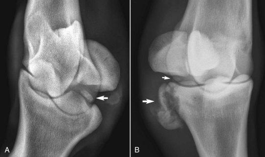
The fragment size that can be easily removed during arthroscopic surgery is limited, and the instrument portal must be enlarged to accommodate such fragments unless ostectomy is performed. The fragment must be located in the joint or at least close to it, a decision that must be made before surgery because arthroscopic surgery is of little benefit if the fragment is not in the joint or close enough to make the approach reasonable and safe. In the metatarsophalangeal joint, large plantar process fragments from the proximal phalanx are often located extraarticularly, and although they may still be best removed using conventional surgical techniques, I have removed large fragments with arthroscopic instrumentation; surgery is facilitated by use of a motorized synovial resector (Figure 23-1). Differentiation should be made preoperatively between fragments free in a joint, which are accessible to surgical removal, and those embedded in the joint capsule and therefore not readily removed. In the stifle joint, fragments in the distal aspect of the femoropatellar joint may appear radiologically as if they are in the femorotibial joint. Rare fragments in the caudal pouches of the medial and lateral femorotibial joints can be difficult to retrieve, especially if the precise location cannot be determined radiologically before surgery. The approaches to the difficult caudal pouches of the medial and lateral femorotibial joints were recently refined.4 Highly mobile fragments in a large joint such as the femoropatellar joint can prove challenging to locate. There is still a limited role for arthrotomy in equine surgery. For example, I still prefer to use a small, medial, middle carpal arthrotomy to repair sagittal slab fractures of the third carpal bone (C3) because I believe I can more accurately place the screw in the medial aspect of the C3, between the C3 and the second carpal bone.5 Others prefer a technique using arthroscopic guidance.1


Jun 4, 2016 | Posted by in EQUINE MEDICINE | Comments Off on Arthroscopic Examination

Full access? Get Clinical Tree

Get Clinical Tree app for offline access