The Stifle

Chapter 46The Stifle


Anatomy


Femoropatellar Joint


The articulation between the patella and the trochleas of the femur forms the femoropatellar articulation. The patella has three straight ligaments. The medial patellar ligament attaches to the medial border and the distal aspect (apex) of the patella through the parapatellar fibrocartilage. Arthroscopically the ligament can be viewed beneath the joint capsule. Distally the medial patellar ligament attaches medial to the groove on the cranial aspect of the tibial tuberosity. The middle patellar ligament originates on the cranial part of the patella just proximal to the apex and inserts in the distal part of the groove of the tibial tuberosity. The lateral patellar ligament extends from the lateral aspect of the patella to the lateral part of the tibial crest. The biceps femoris has a tendon of insertion on this ligament.3 Femoropatellar ligaments also reinforce the joint capsule medially and laterally, the lateral ligament being the more distinct.


The medial trochlear ridge of the femur is larger and more rounded than the lateral trochlear ridge and articulates with the medial part of the patella and the fibrocartilage of the medial patellar ligament. The joint capsule has a large suprapatellar pouch and inserts abaxially on the trochlear ridges forming lateral and medial recesses, the lateral being the smaller. A large fat pad is cranial to the joint capsule, proximal and distal to the patella. In my experience, most horses have a slitlike opening into the respective femorotibial joints at the distal end of the medial trochlear ridge and frequently the lateral trochlear ridge as well. Latex passed from the femoropatellar to the medial femorotibial joint in 60% to 65% of horses and in the reverse direction in 80% of horses.3 Diffusion of mepivacaine between all the compartments may occur in about 75% of horses.4


Femorotibial Joints


The medial and lateral femorotibial joints are separate compartments, which are divided by an intact median septum in a healthy joint. However, they may communicate after trauma.5 The crescent-shaped, fibrocartilaginous medial and lateral menisci lie between the respective femoral and tibial condyles to form a congruent articulation.2 Both are attached to the tibia, cranial to the intercondylar eminences, by cranial ligaments. The medial ligament wraps around the cranial aspect of the medial intercondylar eminence before inserting on the tibia. The medial meniscus is also attached caudally to the medial intercondylar eminence by the caudal ligament, which can be seen arthroscopically in the caudal part of the medial femorotibial joint. The lateral meniscus attaches caudally to the popliteal notch of the tibia and through the strong meniscofemoral ligament to the caudal part of the intercondylar notch of the femur. Only the cranial and caudal poles of the menisci can be viewed arthroscopically because of the close apposition of the tibia and femur, but the respective cranial ligaments are clearly visible. During flexion the menisci slide caudally, and during extension they slide cranially.


The cranial cruciate ligament has its tibial attachment cranial to the medial intercondylar eminence and its femoral attachment in the lateral part of the intercondylar notch. The cranial cruciate ligament lies beneath the median septum and usually cannot be directly viewed arthroscopically without removing the septum. A better view is often gained from the lateral compartment. The caudal cruciate ligament originates in the popliteal notch of the caudal aspect of the tibia and runs proximally, medial to the cranial cruciate ligament, to insert cranially in the intercondylar notch of the femur.6 The ligament can be viewed beneath the septum in the cranial and caudal medial compartment of the femorotibial joint. In the dog, the cranial cruciate ligament is under tension during extension of the femorotibial joint,7 and this may be so in the horse.


The collateral ligaments both originate proximally on the respective epicondyles of the femur. The medial collateral ligament inserts distally on the tibia distal to the medial condyle and has attachments to the medial meniscus. The lateral collateral ligament lies over the popliteal tendon and inserts distally on the head of the fibula. The popliteal tendon originates close to the lateral collateral ligament on the femur and courses distally and caudally, in close apposition to the femoral condyle, to a triangular area of insertion on the proximal caudal aspect of the tibia. The tendon is viewed arthroscopically in the cranial aspect of the lateral femorotibial joint and also in the caudal part, which it effectively divides, limiting the arthroscopic accessibility. The tendon of origin of the long digital extensor muscle can be followed arthroscopically in the cranial compartment of the lateral femorotibial joint from its origin on the extensor notch of the femur and is usually invested within the joint capsule, although in some traumatized joints, the tendon appears separate.5


Diagnosis


Gait and Manipulative Tests


Differentiating stifle lameness in the horse by studying the gait is difficult because the reciprocal apparatus coordinates the movement of the whole limb. In my view, attributing the cause of certain gait changes to stifle pain is not possible. Some horses with stifle pain may carry the stifle slightly abducted, but this is not specificimage. A careful analysis of the whole limb is required to establish the site of pain causing lameness. Other gait changes that may be seen with stifle lameness, but which are also not specific, are a reduced cranial phase of the stride and a reduced flexion of the limb in flight. Many horses with stifle pain dislike going downhill. Horses with delayed release or upward fixation of the patella tend to avoid fully extending the limb and appear to have a crouching gait. Flexion of the upper limb exacerbates lameness in horses with stifle pain, and abduction of the limb may be resented. When performing proximal limb flexion tests, holding the limb at midmetatarsal level, rather than by the foot, helps to differentiate between upper and lower limb pain.


Three specific manipulative tests have been described for the stifle. These are the cruciate test, collateral ligament test,8 and patellar displacement test. Most horses with clinically significant stifle pain resent these tests, which makes the tests difficult to perform and to interpret. All manipulation or flexion tests should be done on the contralateral limb first. For the cruciate test, the affected limb should be weight bearing. The head of the tibia is pushed caudally and then released 5 to 10 times before trotting the horse. Laxity is supposed to be appreciated and lameness exacerbated if severe cruciate injury exists. I have never found this test effective. Pain in the affected joint provokes strong guarding by the horse so that the procedure is impossible to perform. The medial collateral ligament test involves abducting the distal limb against shoulder pressure exerted on the femorotibial joint 5 to 10 times before trotting the horse. Horses with ruptured medial collateral ligaments are so painful and instability is so great that this test is inappropriate, but it can be useful for a sprain of the ligament. The lateral collateral ligament is less often affected, but it can be tested by pulling the distal aspect of the limb medially. Lameness associated with problems with patella release may be worsened by pushing the patella proximally several times with the horse weight bearing before trotting, but again this is often an unrewarding test.


Diagnostic Analgesia


In many horses with low-grade stifle lameness, positive diagnostic analgesia is the only way to localize the site of pain, so it is an important test. Because diffusion of local anesthetic solution between the three joint compartments is so variable,4 all three must be blocked to ensure a valid test. Alleviation of lameness after analgesia of one compartment does not necessarily infer that that compartment is definitely the source of pain.


I use a 5-cm, 19-gauge needle for each joint compartment and up to 30 mL of local anesthetic solution because experience has shown that 20 mL in each compartment might be incompletely effective in a 600-kg horse. Strict aseptic procedure should be followed. Arthrocentesis of the femoropatellar joint is well tolerated and performed first. An intradermal bleb is usually unnecessary. However, an inexperienced veterinarian may find one helpful for the medial femorotibial compartment because some horses are sensitive to injection at this site. My preferred approach for the femoropatellar joint is between the middle and medial patellar ligaments. Synovial fluid is infrequently retrieved from this site unless the joint capsule is distended. If a synovial fluid sample is required, it may be retrieved more easily through a lateral approach.9 The lateral cul-de-sac is entered caudal to the caudal edge of the lateral patellar ligament and 5 cm proximal to the tibial condyle. The medial femorotibial compartment is entered over the medial tibial condyle between the medial patellar ligament and the medial collateral ligament. A small outpouching of the joint capsule may be palpated. The lateral femorotibial compartment is best approached just cranial or caudal to the long digital extensor tendon and close to the tibial plateau. Less space is available between the meniscus and the joint capsule in the latter approach, so the former is preferred. An improvement in lameness can be expected in 30 minutes, but the clinician is wise to allow at least 1 hour for the final assessment.


Horses with a number of conditions causing lameness in the stifle respond incompletely or not at all to intraarticular analgesia. Horses with medial or lateral collateral ligament or patellar ligament injuries may be unaffected. Horses with subchondral bone cysts in the medial femoral condyle show a variable response, ranging from resolution of lameness to little change, and analgesia can take a long time to take effect. Horses with conditions that cause severe lameness are often only partially improved by analgesia; these conditions include infections; fractures, particularly patellar and tibial crest fractures; advanced osteoarthritis (OA); and severe cruciate and meniscal tears.


Imaging Considerations


Radiography


Although many stifle injuries are not associated with detectable radiological changes, radiography is usually the first imaging mode to be used once the site of pain causing lameness has been established as the stifle, or if the distal aspect of the limb has been excluded as a potential source of pain. An x-ray machine capable of producing at least 90 kV and 20 mAs is required. In larger horses, adequate definition will only be achieved with even higher-powered x-ray generators. Fast-screen film combinations can be used particularly for caudocranial images. Using as slow a combination as possible is always worthwhile, commensurate with safe practice, to achieve the best definition on the radiograph. Large cassettes are necessary and should be held in a cassette holder with a long handle. Because of the difficulty of aligning the cassette perfectly in the standing horse, using a grid is impractical, although one can be used if the horse is under general anesthesia. Many horses dislike having cassettes placed close to the stifles, so great care must be taken with this procedure. If any doubt exists about the horse’s temperament, the horse should be sedated.


Five standard images are most commonly used: lateromedial, flexed lateromedial, caudocranial, caudolateral-craniomedial oblique, and cranioproximal-craniodistal oblique (skyline). The radiographic anatomy of the soft tissue attachments of the stifle is well described.10


Ultrasonography


A substantial proportion of stifle lameness is caused by soft tissue damage; therefore ultrasonography has a potential diagnostic role in defining the injury and has several advantages over other imaging diagnostic techniques. At present, ultrasonography is the only method of assessing soft tissue injury in the stifle in a standing horse. The disadvantages are that ultrasonography requires experience to be used effectively and a good-quality ultrasound scanner with a sector and a linear array transducer is necessary. Transducer frequencies of 7.5 and 5 MHz are needed to image the cranial aspect of the stifle, but a 3-MHz transducer is required to image the caudal part of the stifle.12-15 When it becomes available, magnetic resonance imaging (MRI) is likely to be a superior imaging technique for soft tissues of the equine stifle.16 Computed tomography (CT) has already proven useful for evaluation of meniscal tears and for detection of cartilage and subchondral and trabecular bone injury.17


Ultrasonography can be valuable for differentiating joint capsule distention from extraarticular swelling. Soft tissue structures that can be imaged include the patellar ligaments, the menisci and the respective cranial ligaments, the collateral ligaments, the cranial and caudal cruciate ligaments, the meniscofemoral ligaments, the origin of the long digital extensor tendon, and the popliteal tendon. The articular cartilage and bony outline of the femoral trochlear ridges and the cranial and caudal third of the femoral condyles may also be imaged.12-15


The patellar ligaments and the collateral ligaments can be imaged longitudinally and transversely with a 7.5- to 10-MHz linear array transducer with the horse weight bearing. The middle patellar ligament is the most obvious of the three and is a useful landmark. The femorotibial collateral ligaments can be imaged from the attachments on the distal lateral or medial femoral epicondyles to the attachments on the proximal medial or lateral aspects of the tibia, and they lie over the respective menisci.18 The menisci can also be imaged with this transducer from caudal to the medial and the lateral patellar ligaments. They appear as wedge-shaped structures of moderate echogenicity with the base of the wedge closer to the transducer. The cranial ligaments of the menisci are more easily imaged with a small convex array or sector transducer, which can be aimed more perpendicular to the meniscal ligaments and which can be more easily positioned between the patellar ligaments. The cruciate ligaments are difficult to image because aligning the transducer perpendicular to the fibers of the ligament is difficult. Only a small length of ligament can be imaged at a time, which can make interpretation equivocal. Cruciate ligaments can only be viewed with the stifle in a flexed position using a convex array or sector scanner. Useful information on the surfaces of the femoral trochlear ridges and condyles can be obtained. The condyles are imaged with the stifle in a flexed position, and this can be helpful in diagnosing subchondral bone cysts.19 From caudal, a sector transducer is preferable for imaging the caudal cruciate ligament and the meniscofemoral ligaments, and a 3-MHz transducer is necessary in large horses.13


Scintigraphy


Scintigraphy has been used on horses for more than 30 years, but little published evidence assesses its specificity and sensitivity for conditions of the stifle. A recent study of 16 horses indicated moderate to high sensitivity of scintigraphy for detection of meniscal damage, cruciate ligament injury, or articular cartilage pathology in the stifle using arthroscopy as the gold standard, but specificity was low, indicating a high risk of false-negative results.20 For most stifle conditions, scintigraphic findings are variable, and although positive findings are obviously helpful, negative findings can also add useful information. The clinical significance of any scintigraphic result should be confirmed as exhaustively as possible by other tests, particularly diagnostic analgesia because false-positive results also occur. In the normal adult stifle, the caudal aspect of the tibial epiphysis often has highest uptake of radiopharmaceutical.21


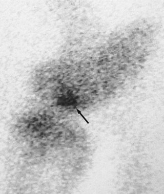
Nuclear scintigraphy is most consistently valuable in diagnosing incomplete avulsion fractures associated with the stifle.22,23 However, although positive results may be obtained within 24 hours of injury, in some horses at least 3 days must elapse before clinically significant increased radiopharmaceutical uptake (IRU) occurs. Subchondral cystic lesions that cause lameness may be scintigraphically positive or negative (Figure 46-1). The lesions are more likely to be detected in older horses, using a caudal image, than in an immature horse with high background activity. Absence of radiopharmaceutical uptake was thought to be the result of osteoclasts being the dominant cells in certain stages of the condition.24,25 However, in the Editors’ experience the majority of subchondral cystic lesions do have focal IRU. Although scintigraphy has been used to assess osteochondrosis in people,26 bone scan findings are not well documented in horses. I have seen positive and negative scintigraphic results associated with clinically significant osteochondrosis of the lateral femoral trochlea. Soft tissue injuries of the stifle are often scintigraphically negative, but in my experience IRU can occasionally be encountered in these horses, especially in association with enthesopathy.


Scintigraphy is certainly a useful tool as a diagnostic aid for lameness in the stifle and can be useful for evaluating the bone activity of lesions. Occasionally horses may respond positively to intraarticular analgesia, but no clinically significant abnormalities are found on radiography, ultrasonography, and arthroscopy. IRU in the stifle may be the only finding. Making a definitive diagnosis in these horses is difficult, although conceivably subchondral bone pain may be a possible cause of the lameness. One should bear in mind that many stifle conditions are negative scintigraphically. Conversely, the joint may be scintigraphically positive when the cause of lameness is elsewhere in the limb.


Articular Diseases


Femoropatellar Joint


Osteochondrosis


Osteochondrosis has been recognized in horses for more than 50 years and is an important cause of stifle lameness in young horses. The exact cause of the disease is still not well defined, but several factors are known to influence its development. Adequate dietary copper is important. Mare’s milk is relatively low in copper, and because the foal relies on copper stored in its liver during late pregnancy, it will be deficient if the mare’s diet contains insufficient copper. The zinc/copper ratio is also important because zinc inhibits the absorption of copper.27 Foals on high-energy diets are more prone to the disease.28 Insufficient or excessive exercise and trauma may be factors that influence the development of the disease. Genetic factors may predispose to osteochondrosis in the hock of Swedish Standardbreds (STBs).29 Large, fast-growing males are more susceptible. Osteochondrosis is most frequently seen in Thoroughbreds (TBs) and Warmbloods. The lesions probably develop in the first 7 months of life,30 but sometimes clinical signs may not be manifest until the horse is brought into work. In the sports horse this may be as late as 5 years of age or even older when a mild to moderate lameness may develop as the horse begins more serious work. In my experience, surprisingly severe lesions can remain undetected until this time. Lesions are most commonly seen on the lateral trochlear ridge of the femur31 but do occur on the medial trochlea, intertrochlear groove, and patella and are often bilateral.


Signs and Diagnosis


Osteochondrosis may be present asymptomatically. Lameness is often acute in onset and varies from a subtle gait deficit to marked lameness. Lameness is often more acute in onset and more severe in foals and young yearlings than in older horses. Distention of the femoropatellar joint capsule is often present and can be severe, especially in foals and yearlings. Gluteal muscle atrophy is seen in horses with severe lesions. Flexion tests are mostly positive. Synovial fluid may be hemorrhagic, but it is often normal. Intraarticular analgesia of the femoropatellar joint should improve lameness. However, in mature horses lameness may be mild, with no effusion and a poor response to intraarticular analgesia. In such horses arthroscopic evaluation often reveals an extensive area of cobblestone-like cartilage, with extensive fibrillation over a large portion of the lateral trochlea of the femur. This cartilage is often well adherent to the subchondral bone and little clinical benefit is derived from surgery.


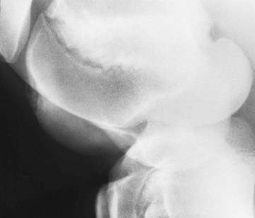
Radiological changes include the following: no detectable signs, slight loss of contour or loss of outline of the lateral trochlear ridge, irregular defects in the trochlear ridge, radiodense fragments within the defect (Figure 46-2), round radiopaque bodies loose in the joint, and more rarely, irregularities on the patellar apex or the medial trochlear ridge.1,32,33 Lateromedial and caudolateral-craniomedial oblique images are the most useful projections. Horses with radiological lesions do not always show lameness.25,27 Some horses have no detectable radiological abnormalities, but osteochondrosis lesions are diagnosed on arthroscopic examination. Lesions without fragmentation in young foals may resolve with time,32,33 but many progress.34


Treatment


Conservative management, with confinement and correction of dietary imbalances, and arthroscopic debridement of the lesions are the main treatment options. Conservative management is appropriate for horses with mild lesions. Because of the insensitivity of radiological evaluation of osteochondrosis lesions, an argument can be made for arthroscopic examination of most horses with lesions seen radiologically or persistent femoropatellar effusion and lameness that fails to respond to conservative treatment.35 Arthroscopic examination would ensure that horses with radiologically silent cartilage lesions are treated effectively. OA can also be evaluated. For horses with fragmentation and clinically significant radiolucent defects of subchondral bone, arthroscopic surgery is probably the treatment of choice.31 In my opinion, more caution is required when treating foals arthroscopically because some foals with large lesions seen radiologically are found to have intact cartilage at arthroscopy. Debridement of such lesions can leave unacceptably large deficits in the lateral trochlear ridge. Reattaching these cartilage flaps with polydioxanone pins may be a more appropriate treatment and has shown good results with seven of nine foals achieving athletic use.36


A variety of lesions are seen arthroscopically. The articular cartilage may appear intact, but if probed, large flaps may lift from the subchondral bone. Large defects on the lateral trochlear ridge may contain nodules of mineralized cartilage, which are readily removed, and often large tufts of fibrillated cartilage are associated with the lesion. The exposed subchondral bone is frequently soft and crumbly. Extensive areas of fibrillated cartilage over the adjacent lateral trochlear ridge and the opposing patella are often present in older horses.5 Loose cartilage should be removed, and the lesion should be curetted to healthy subchondral bone, which is a source of pluripotential cells. The cartilage perimeter should be vertical to allow better attachment of tissue regrowth.37 Thorough lavage to remove all debris, particularly of the suprapatellar pouch, at the completion of surgery is essential. Six weeks of stable rest with daily handwalking after 2 weeks is recommended postoperatively. The horse should then be left for another 4 to 5 months at pasture before returning to work.


Jun 4, 2016 | Posted by in EQUINE MEDICINE | Comments Off on The Stifle

Full access? Get Clinical Tree

Get Clinical Tree app for offline access