2 The Oral Examination and Disease Recognition
When you have completed this chapter, you will be able to:
• Describe the normal occlusion seen in dogs and cats.
• Differentiate between pedodontics, orthodontics, periodontics, prosthodontics, and endodontics.
• Define anodontia and list possible conditions with which it is associated.
• Describe the three classes of malocclusion and discuss the various clinical presentations seen with these conditions.
• List and describe the classifications of oropharyngeal inflammation.
• Describe possible abnormal conditions of the tooth surface, including abnormalities in enamel formation.
• List and describe the various fracture classification schemes that apply to veterinary dentistry.
• List and describe common oral medical diseases, including neoplastic conditions of the oral cavity.
Canine oral viral oral papillomatosis
Contact mucositis (also contact mucosal ulceration)
Cranial mandibular osteodystrophy
Mandibular periostitis ossificans
Maxillary-mandibular asymmetry
Peripheral odontogenic fibroma
Ulcerative eosinophilic stomatitis
The wide variety of genetics in the dog (and to a lesser degree the cat) gives rise to a wide variety of size and developmental abnormalities. Figure 2-1 illustrates this variety in the difference in size between a Chihuahua and Saint Bernard.
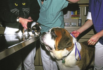
FIGURE 2-1 The differences in the canine size expressed by looking at a Chihuahua and Saint Bernard.
Oral Examination
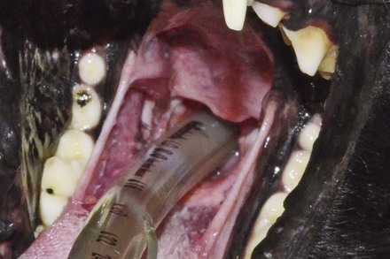
The duties of veterinarians and their staff are described in the practice acts of each state. Because state law varies, veterinary staff members should refer to the specific practice acts of the state in which they are employed. However, some generalities apply. The oral examination should always be conducted in a systematic manner. In many states, it is legal for the registered veterinary technician to induce anesthesia under orders of a licensed veterinarian. The placement of the endotracheal tube is a good opportunity for visualization of the pharynx, tonsils, and tongue (Figure 2-2). While scaling and polishing the teeth, the technician can closely observe the tooth surface and gingiva. The technician should always note any abnormalities and report them to the doctor.
Normal Occlusion
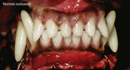
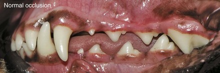
Normal occlusion in dogs and cats is a scissors bite, in which the mandibular (lower) teeth come into contact with the palatal side (inside) of the maxillary (upper) teeth (Figures 2-3 and 2-4). Normally, the cusp of the mandibular incisors rests on a ledge on the palatal side of the maxillary incisors known as the cingulum. The mandibular canines fit in the diastema (space) between the lateral incisor and maxillary canines. The cusp of the mandibular first premolar fits midway between the maxillary canine and first premolar. The remainder of the premolars are intermeshed in a similar fashion: The lower teeth are approximately one-half a tooth in front of their maxillary counterparts.
Specialties of Dentistry
In the past, veterinarians were expected to treat all animals for any conditions they might have. Veterinary medicine has since become more sophisticated; with the advancement of knowledge, no veterinarian can perform all facets of veterinary medicine. Consequently, the field is now increasingly specialized. In fact, human dentistry is subdivided into specialties (Table 2-1), a phenomenon that is becoming increasingly common in veterinary dentistry as well. The more a practitioner performs a particular skill, the better the practitioner becomes. Therefore treatment of advanced periodontal disease, fractured teeth, and orthodontic conditions may not be in the patient’s best interest when the practitioner performs these procedures only occasionally. Furthermore, preparing the practice for the occasional specialized procedure requires considerable expense, training, and time. In this case, the “win-win” solution is referral.
| Branch of Dentistry | Area of Specialization |
|---|---|
| Endodontics | Treatment of diseases that affect the tooth pulp and apical periodontal tissues |
| Exodontics | Extraction of teeth and related procedures |
| Oral surgery | Surgery of the oral cavity |
| Orthodontics | Guidance and correction of malocclusion of the juvenile teeth and adult tooth positioning |
| Periodontics | Study and treatment of diseases of the tooth-supporting tissues |
| Prosthodontics | Construction of appliances designed to replace missing teeth and/or other adjacent structures |
| Restorative/operative dentistry | Restoration of form and function of teeth |
From Wiggs RB, Lobprise HP: Veterinary dentistry: principles and practice, Philadelphia, 1997, Lippincott-Raven; Holmstrom SE, Frost P, Eisner ER: Veterinary dental techniques, Philadelphia, 1998, WB Saunders.
Oral Diseases and Dental Specialties
Pedodontics
Puppies and kittens exhibit a variety of dental conditions, both genetic (inherited) and acquired.
Missing teeth
Anodontia refers to the absence of teeth. Teeth may be missing because they never developed in the first place, are slow to erupt, or were present and fell out. Dental radiographs must be taken to evaluate the area of the missing tooth. In dental charts a circle around the tooth indicates that it is missing. A radiograph should be taken to evaluate the oral structure for missing teeth (see Chapter 11 for technique). The radiograph indicates whether the root is missing or retained. The absence of the root may be inherited or the result of trauma.
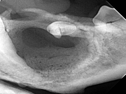
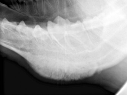
Some breeds, notably the Boxer, pug, and dachshund, are prone to retaining their teeth in the bone subgingivally. This may lead to the formation of a dentigerous or follicular cyst. The cyst is lined by epithelial cells derived from the enamel epithelium of the tooth-forming organ. Figure 2-5 shows a radiograph of a cyst in a 5-year-old Boxer.
Persistent primary teeth
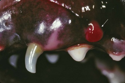
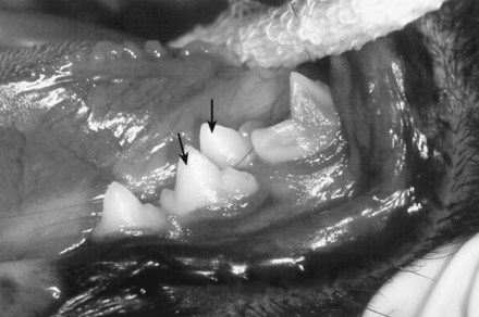
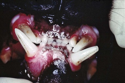
Persistent primary (also called retained, deciduous, or baby) teeth may cause orthodontic and periodontic abnormalities. Extraction of these teeth may help prevent such complications. In the patient shown in Figure 2-6, the primary tooth is displacing the maxillary canine. In addition to causing the possible displacement of the adult canine, the abnormal periodontal border may cause periodontal disease resulting from plaque being trapped between the primary and adult teeth. The general rule applies: There is no room for two teeth of the same type in the same mouth at the same time. Unless they are extremely loose, retained primary teeth should be extracted as soon as possible after the adult tooth starts to erupt. The patient in Figure 2-6 has a persistent left maxillary canine (1c, 604), which should be extracted. The patient in Figure 2-7 has a persistent left maxillary lateral incisor (1i, 603). The important question to answer for this patient is: “where is the adult lateral incisor?” Intraoral radiographs are indicated to see if the adult tooth never formed or if it is impacted and potentially causing a dentigerous cyst.
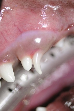
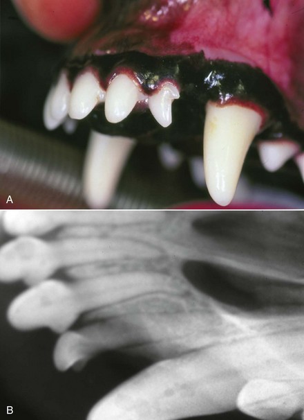
FIGURE 2-7 A, A persistent deciduous lateral incisor. B, Radiograph showing that the adult tooth never formed.
Cranial mandibular osteodystrophy
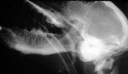
Cranial mandibular osteodystrophy is an inherited condition that occurs primarily in West Highland white terriers and occasionally in other breeds. Nonneoplastic bone forms in the region of the temporomandibular joint (TMJ) and occasionally extends into the mandible. Patients with cranial mandibular osteodystrophy are treated symptomatically for pain, which usually lessens as the patient gets older. Figure 2-8 shows a nonneoplastic bone production in the region of the TMJ.
Mandibular periostitis ossificans
Mandibular periostitis ossificans occurs in immature large breed dogs. It causes a unilateral swelling of the ventral portion of the mandible. It is diagnosed radiographically by a two-layered (double) ventral mandibular cortex. This is periosteal new bone formation, which is thought to be an inflammatory condition that spontaneously disappears (Figure 2-9).
Fractured deciduous (primary) teeth
Fractured deciduous teeth occur fairly frequently. They may be caused by running into objects, catching rocks or other hard substances, or overzealous playing of games such as tug-of-war. If left untreated, fractured primary teeth may result in abscessation, which can in turn cause a defect in enamel production known as enamel hypoplasia and may or may not form a fistula. Figure 2-10 shows a fractured left maxillary canine (1c, 604). Note the fistula formation (arrow) above the premolar as an extension of the fracture and subsequent abscess. This tooth should be extracted.
Supernumerary teeth
Supernumerary teeth are primarily incisors, although all types of teeth may be supernumerary. One problem that supernumerary teeth may cause is crowding. The patient in Figure 2-11 has a supernumerary left mandibular fourth premolar (4P, 308). The patient in Figure 2-12 has a supernumerary canine tooth.
Peg teeth
Peg teeth are abnormally formed supernumerary teeth. They generally occur in the canine and incisor regions. The treatment is extraction. The patient in Figure 2-13 has a peg tooth located lingual to the left maxillary central incisor (1I, 201).
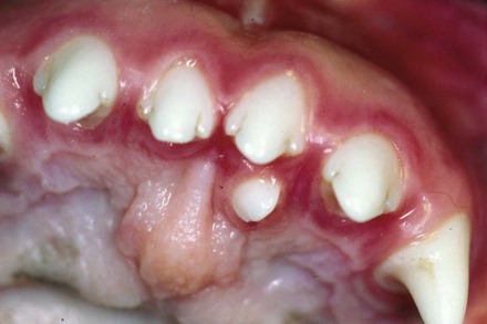
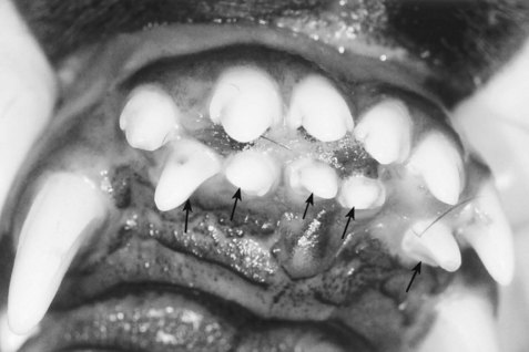
Third set of teeth
Supernumerary teeth may also result from the formation of a third set of teeth. The primary teeth of the patient in Figure 2-14 fell out at the normal time. The outermost row of teeth are adult teeth. A supernumerary third row of teeth is present in this patient. The innermost row of teeth was extracted.
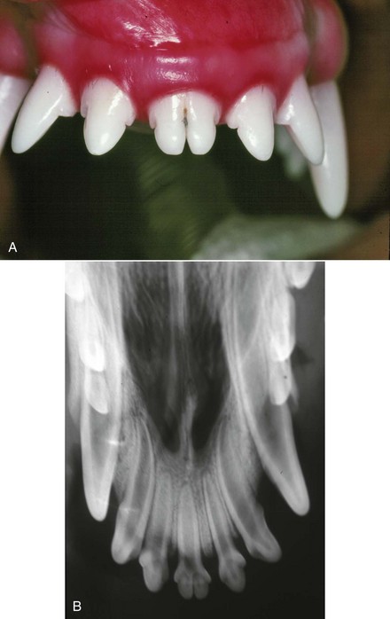
Fused and gemini teeth
Fusion is the joining of two developing teeth that have different tooth buds. A gemini tooth is one in which a tooth bud has partially divided in the attempt to form two teeth. The left central incisor (1I, 201) in Figure 2-15 is a gemini tooth. A radiograph is necessary to differentiate between the two. Two roots will be seen with fused teeth, whereas only one root with twin crowns will be seen with gemini teeth.
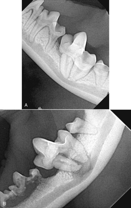
Dilacerated roots
A dilacerated root is an abnormally formed root, which may be caused by trauma during the tooth’s development or by genetic conditions. It may be an unusual finding discovered while taking dental radiographs without pathology or it may be accompanied by severe pathology. The patient in Figure 2-16, A, has congenital dilacerations of the mandibular first molars. Treatment was recommended at the time of the discovery, but the client declined. Figure 2-16, B, shows the same patient 4 years later. A severe lytic area around the dilacerated roots has developed. The teeth were extracted.
Orthodontic Disease
Class I malocclusions
Spearing (lancing) canines
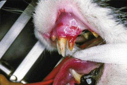
When the maxillary canines are tipped in a rostral position, they are trapped by the mandibular canines. This Class I orthodontic condition is known as spearing canines. Other terms that are sometimes used are lancing and tusk teeth. The condition appears to be genetic and is most prevalent in shelties and Persian cats. Treatment includes orthodontic correction and extraction. The cat in Figure 2-17 has bilateral spearing canines (1C1, 104, 204).
Stay updated, free articles. Join our Telegram channel

Full access? Get Clinical Tree


