1 Introduction to Veterinary Dentistry
When you have completed this chapter, you will be able to:
• Differentiate between the terms mesaticephalic, brachycephalic, and dolichocephalic.
• Identify the anatomic components that comprise the mandible and maxilla.
• Describe the structure of the teeth and supporting tissues.
• List the dental formulas for dogs and cats.
• Identify the terms used to designate position and direction in the oral cavity.
• Describe the anatomic and Triadan numbering systems.
• Describe the method for recording pathology on a dental chart.
General Anatomy
Types of Heads
Mesaticephalic
Mesatic means medium. Mesaticephalic is the most common head type. Poodles, corgis, German shepherds, Labrador retrievers, and domestic shorthair cats are typical examples (Figure 1-1).

Brachycephalic
Brachy means short. Brachycephalic animals have short, wide heads. This characteristic commonly results in crowded and rotated premolar teeth, a condition that may lead to periodontal disease. Boxers, pugs, bulldogs, and Persian cats are common examples of the brachycephalic type (Figure 1-2).


Maxilla
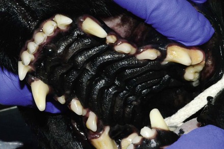
The upper jaw, called the maxilla, is made up of many bones (Figure 1-4). The incisal and maxillary bones hold the teeth. The roof of the mouth comprises the hard and soft palates. The hard palate is the portion of the roof of the mouth that consists of hard bone. The hard palate is covered with a mucous membrane that has irregular ridges, called the rugae palatinae. The incisive papilla lies behind the central incisors. The nasopalatine ducts exit on each side of the incisive papilla. The soft palate is the posterior portion of the roof of the mouth, which does not have underlying bone. This portion separates the oral cavity from the pharynx, which leads to the nasal cavity. Close observation reveals that the teeth are surrounded by the gingiva. The area in which the two jaws join in the back of the oral cavity is known as the lateral palatine fold.
Mandible
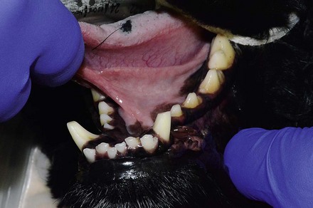
The lower jaw is known as the mandible (Figure 1-5). It is connected to the maxilla by a hinge joint called the temporomandibular joint (TMJ). The two mandibles are fused together at the mandibular symphysis. The tongue lies between the two mandibles, and the structures and surfaces beneath the tongue are referred to as sublingual. The mandible is covered ventrally by muscle and skin. The oral cavity is covered with a mucous membrane, which becomes the gingiva at the mucogingival line.
Tooth Anatomy
External Tooth
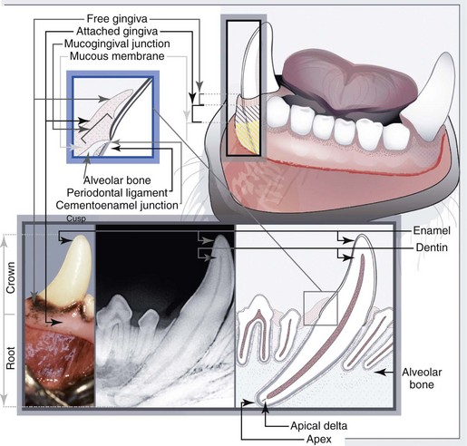
The tooth may be divided into the crown, neck, and root (Figure 1-7). The tip of the crown is known as the cusp. The crown is covered with enamel, the hardest substance in the body. It will survive normal use and even some abuse without problems. However, it may fracture in patients who chew bones and other hard substances. Normally, enamel is present only above the gumline. Enamel is produced by cells called ameloblasts as the tooth is developing. Where the enamel thins close to the gumline, many teeth have a slight indentation. This indentation is known as the neck. Underneath the gumline is the root. The deepest part of the root is known as the apex. At the apex, blood vessels and nerves enter the tooth through a series of small channels known as the apical delta or through larger canals known as the apical foramen. The cusp is the tip or pointed prominence on the occlusal surface of the crown.
Stay updated, free articles. Join our Telegram channel

Full access? Get Clinical Tree


