The Opportunistic Mycoses

Chapter 48 The Opportunistic Mycoses


Infection is a rare consequence of environmental exposure of animals to normally nonpathogenic fungi. Natural resistance to most of these agents is high, and most are of low virulence. However, agents of low virulence may act as pathogens when animals become stressed or otherwise debilitated, are treated with antibiotics, or following breakdown of the mucocutaneous barrier. Opportunistic fungi (a categorization without taxonomic standing) take advantage of these situations and cause infections. Most opportunistic mycoses are attributed to Candida and Aspergillus spp., and members of the class Zygomycetes.


THE GENUS CANDIDA


The genus Candida belongs to the family Cryptococcaceae (order Cryptococcales, class Deuteromycetes) and contains more than 200 species. Taxonomic relationships within the genus are poorly defined. They reproduce by budding and production of chlamydospores. Pseudohyphae may be absent, present in rudimentary form, or well developed. Hallmarks of the genus are failure to produce acetic acid, ballistoconidia, or arthrospores, and lack of red or orange pigmented colonies.


Diseases and Epidemiology


Candida spp. are normal inhabitants of the alimentary, upper respiratory, and genital mucosae of animals, and primarily associated with infections of the skin and mucous membranes; however, they can invade nearly every organ in the body. Disruption of the epidermal barrier by trauma, antibiotic therapy, inflammation, immunosuppressive disease, or glucocorticoid therapy may allow the yeasts to overcome the normal cutaneous defense mechanisms.


Human infection often follows some sort of debilitation, such as hormone imbalance or application of immunosuppressive therapy. Infections are common in patients with acquired immunodeficiency syndrome (AIDS), and intravenous drug users are subject to endocarditis. Foreign bodies (such as catheters) can promote colonization, and hyperalimentation can predispose to infection.


Cutaneous candidiasis occurs infrequently in dogs and cats. In the dog, infection has been associated with Candida albicans, Candida parapsilosis, and Candida guilliermondii. Exfoliative dermatitis may be found on the muzzle, inguinal area, scrotum, and dorsal and lateral aspects of the feet. In addition, seborrhea, pruritus, and alopecia may be noted. Candida albicans has been associated with an outbreak of otitis externa in a pack of foxhounds, and localized and disseminated infections have been reported in cats. Many of the cats were concurrently infected with feline panleukopenia or feline immunodeficiency virus.


Gastroesophageal candidiasis, in association with gastric ulceration, has been described in foals and calves. Animals present clinically with signs of colic that are nonresponsive to medical treatment. Candida albicans or Candida krusei has been recovered from cultures of gastric fluid. Stress is an important predisposing factor.


Crop mycosis (“thrush”) of poultry is caused by C. albicans, and serious outbreaks have been reported in many species of birds. The crop, mouth, esophagus, proventriculus, and gizzard are most frequently affected, and lesions consist of white plaques or pseudomembranes adherent to the mucosal surfaces (Figure 48-1, A). Superficial ulcers occur when plaques slough, and mortality may be high in young birds. Disease is frequently associated with other debilitating conditions such as intestinal coccidiosis or unsanitary, crowded housing.
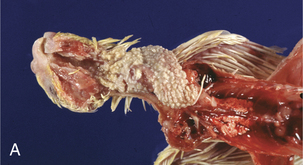
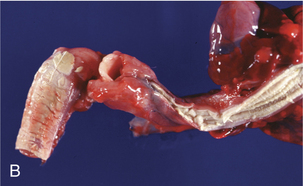
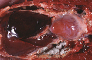


The agent of candidiasis in swine is also C. albicans, and disease is associated with stress or immunosuppression and unsanitary housing conditions. Cutaneous and mucocutaneous disease has been described. Circular skin lesions are covered with thick, gray exudates, and alopecia may be apparent. In chronic infections the skin may become wrinkled and thickened. The lesions of mucocutaneous disease are similar to those observed in poultry, and are characterized by pseudomembrane formation. Similar gross lesions can be found in the javelina (Figure 48-1, B).


Pathogenesis


Depression of cell-mediated immunity is likely an important factor in pathogenesis of candidiasis. Prolonged immunosuppression, neutropenia associated with chemotherapy, diabetes mellitus, and long-term glucocorticoid or antimicrobial treatments are predisposing factors. However, epithelial colonization can occur even in immunocompetent hosts, especially when an epithelial breach occurs. Virulence attributes include factors that mediate adherence to epithelial and endothelial cells, extracellular enzymes, and phenotypic switching.


Several types of adhesive interactions have been described. Candidal surface protein recognition of host-cell–surface carbohydrate results in a lectinlike interaction. Four structurally related adhesins, members of a class of glycosylphosphatidylinositol-dependent cell wall proteins, mediate attachment. Invasion of mucous membranes appears to follow adhesion of the yeasts to a debilitated surface, with subsequent development of pseudohyphae that invade the superficial epithelium.


Production of extracellular cytotoxic phospholipases and proteases correlates with virulence. Blastospores develop hyphae with phospholipase activity concentrated at their growing tips. This facilitates tissue invasion because host-cell membranes are injured as the yeasts germinate. Isolates that adhere most strongly to buccal epithelial cells and are most virulent for mice produce high levels of extracellular enzymes. Phospholipases, in particular, promote adherence of yeasts to host cells. Production of aspartyl proteinase facilitates penetration of keratinized epithelia.


Phenotypic or phase switching is a gene-regulated process whereby Candida spp. make the transition from budding to hyphal forms. Hyphae are commonly thought of as the invasive form, whereas the yeast form is responsible for colonizing epithelia. Tissue penetration and dispersal are mediated by a switch to the hyphal form. This process further enables the fungus to adapt to hostile environmental conditions. Phenotypic switching is accompanied by changes in antigen expression, perhaps allowing the organism to evade the host immune response.


Diagnosis


Isolation of Candida spp. from clinical specimens does not confirm candidiasis as the diagnosis. The yeasts must be demonstrated either in a direct examination of lesion material or in tissues through histologic evaluation.


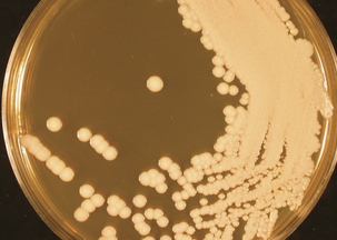
Candida spp. grow readily on a variety of nonselective fungal agars, such as Sabouraud dextrose or potato dextrose agars. Colonies are formed within 2 to 3 days at 25° to 30° C and are creamy, white, opaque, and circular (Figure 48-2). Yeast identification is based on microscopic morphology of organisms cultivated on cornmeal-Tween agar; on production of pigment, germ tubes, and urease; and assimilation and fermentation of carbohydrate.


Presumptive differentiation of C. albicans from other Candida spp. involves the germ tube test. The test is performed by placing several colonies from a nonselective fungal medium in animal serum and incubating at 37°C for 3 hours. Microscopic examination reveals short hyphal segments without constrictions, at the junction of the blastoconidium and the germ tube, in positive strains (Figure 48-3). Not all C. albicans strains produce germ tubes, so false negatives may occur.
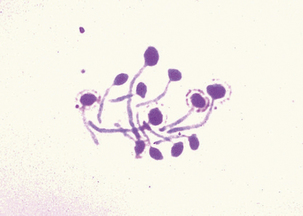


Most Candida spp. can be differentiated through conventional or commercially available biochemical tests, or by molecular methods. Many commercial rapid tests are based on detection of preformed enzymes and provide presumptive identification in as little as 4 hours. Such tests are applicable only to the common yeasts. Molecular techniques are usually practiced only in mycology reference laboratories.


THE GENUS ASPERGILLUS


Aspergilli were first described in 1729 by Micheli, a priest and botanist who thought that the chaining spores of these fungi resembled the brush used for sprinkling holy water, the “aspergillium.” The genus contains more than 180 species and taxonomically is placed in the phylum Ascomycota, order Eurotiales, and family Trichomaceae.


Most cases of animal disease are attributed to Aspergillus fumigatus, Aspergillus flavus, Aspergillus nidulans, and Aspergillus niger. These fungi are ubiquitous in the environment and commonly isolated from plant debris and soil. Aerosol exposure to Aspergillus conidia is constant and inevitable.


Disease and Epidemiology


Human aspergillosis in the United States is the second most common fungal disease requiring hospitalization (after candidiasis). Single isolations of Aspergillus spp. from humans may be insignificant, but multiple positive cultures from immunocompromised individuals suggest invasive aspergillosis. Risk factors include leukemic granulocytopenia, corticosteroid or other drug therapy, smoking marijuana, and posttransplant neutropenia.


Manifestations of aspergillosis in animals include mycotic pneumonia, guttural pouch mycosis, chronic rhinitis, systemic disease, cutaneous disease, allergy, abortion, gastrointestinal aspergillosis, mastitis, and keratomycosis.


Fungi are introduced into poultry houses through spore-contaminated feed or litter. Fungi multiply rapidly under conditions of high humidity, and massive spore inhalation results in acute outbreaks of severe disease associated with high morbidity and mortality. Aspergillosis is sometimes referred to as “brooder pneumonia.” Early lesions consist of small, caseous, white to gray nodules in the lungs or air sacs. Rapid fungal proliferation may line the air sacs with characteristic green hyphae. Acute aspergillosis occurs mainly in poults or chicks, and affected birds are dyspneic, anorexic, and febrile; death is rapid, usually within 48 hours. Chronic disease is seen in older birds, with clinical signs such as coughing, sneezing, ataxia, torticollis, corneal opacity, and emaciation. Trachea and bronchi may be filled with caseous exudate, and white to yellow circumscribed nodules may be visible in the brain. Although the respiratory system is the primary target, other manifestations include dermatitis, osteomycosis, ophthalmitis, enteritis, and encephalitis (Figure 48-4). Aspergillus fumigatus and A. flavus are the etiologic agents most often recovered from lesions.


Horses experiencing enteritis, typhlocolitis, and other diseases of the gastrointestinal tract can be predisposed to pulmonary aspergillosis. Inhalation of spores from moldy feed is probably the primary route of exposure, but cases have also been associated with carriage of spores to lung by migrating larval parasites.


Mycosis of the equine guttural pouch (auditory tube diverticulum) is a sporadic life-threatening infection characterized by necrotizing inflammation with formation of a diphtheritic membrane within the pouch. In most instances it is caused by Aspergillus spp. Infection begins on the roof of the pouch, and fungi proliferate and invade the internal carotid artery and vein and the glossopharyngeal nerve. Fatal hemorrhage follows erosion of the artery. Common clinical signs are epistaxis, dysphagia, parotid pain, abnormal head position, and unilateral mucopurulent nasal discharge. Endoscopic examination of the guttural pouch reveals a white diphtheritic membrane. Peak incidence is during the summer months, and stabled horses are at greater risk. Age, sex, and breed are not risk factors.


Canine rhinitis or nasal aspergillosis is a common disease that primarily affects previously healthy, young to middle-aged dolichocephalic and mesaticephalic breeds. Aspergillus fumigatus is the most frequently isolated species. Affected animals sneeze and have a persistent nasal discharge that is nonresponsive to antibiotics. The disease tends to be invasive and may involve the paranasal sinuses as well as the nasal cavity. Destruction of the turbinate bones with penetration into the brain may result. Immunosuppression is a consequence, rather than a predisposing condition, of infection in dogs. Golden retrievers and collies may be at greater risk than other breeds. Other risk factors are exposure to large numbers of spores and previous facial trauma.


Systemic aspergillosis has been reported in horses but rarely in dogs. Systemic or disseminated aspergillosis is seen almost exclusively in German shepherds and is not associated with prior upper respiratory disease.


Canine cutaneous disease with A. niger and Aspergillus terreus is uncommon and poorly characterized. Cutaneous lesions may result either from an extension of systemic disease or through inoculation during skin trauma. Immunodeficiency is an apparent predisposing factor in humans because disease seems to be most common in burn victims, neonates, transplant recipients, and individuals with cancer and AIDS.


Allergic disease results from an overexuberant immune response in a hypersensitive or atopic individual. Heaves in horses is a good example, and the recently described feline asthma is also associated with hypersensitivity to aspergilli. Clinical signs arise from responses in the upper respiratory tract, and include coughing and exercise intolerance.


Aspergillus spp. cause sporadic abortion in cattle and horses. Prevalence of bovine mycotic abortion has been estimated at 2% to 20%, which varies by geographic location and climate; rates are higher in colder regions and during winter, coincident with feeding of hay. Incidence may approach 10% in some herds. The primary lesions of mycotic abortion are in the placenta, which is generally thickened and leathery, with necrotic areas and fungal growth between the cotyledons. Fungi may invade the fetus and produce ringwormlike lesions (which are fungal colonies) on the skin surface. The fetus is usually expelled soon after death. Aspergillus fumigatus is recovered most frequently.


Mycotic gastritis is common in ruminants. Aspergillus spp., as well as zygomycetes, are usually recovered. Young calves are at greatest risk, and the clinical picture includes inappetance, rumen stasis, and pasty, scant, or loose feces. Acute necrohemorrhagic rumenitis and abomasitis are common.


Keratomycosis occurs more frequently in horses than in other domestic animals. Many different fungi are recovered from these infections, but Aspergillus spp. are the most common etiologic agents. Predisposing factors include trauma, corneal disease or surgery, and topical therapy with antibiotics or corticosteroids. In the affected animal, the cornea appears opaque and ulcerated. Endothelial plaques, which are characteristic of this infection, are located in the center of the cornea.


Pathogenesis


Healthy animals and birds can withstand substantial exposure to Aspergillus spores under natural conditions. Inhalation of massive numbers of spores, as may occur when feed or litter is heavily contaminated, may result in infection. Spores are an ideal size for alveolar disposition.


No single, true virulence factor has been identified, and the mechanisms responsible for protective immunity against Aspergillus spp. have yet to be elucidated. Proteases and other toxins may be involved in the pathogenicity of Aspergillus spp. The predominant protease is an elastase, which may cause lung damage by degrading structural barriers within the lung and facilitating invasion. Elastase-negative strains are relatively avirulent in a mouse model of invasive pulmonary disease. Extracellular proteinases of Aspergillus ochraceus are fibrinolytic and anticoagulative, and may explain the apparent affinity of aspergilli for blood vessels. Rapid release of toxic products may be a critical virulence factor in human infections, with most pertinent effects on macrophages. Gliotoxin, produced by some species, inhibits phagocytosis and T-cell activation and proliferation in vitro.


Equine systemic aspergillosis develops in concert with profound neutropenia associated with colitis. Aspergillus spp. invade the disrupted intestinal mucosa, and mycotic emboli disseminate to internal organs and brain. In horses with normal mucosal integrity, predisposing factors may be prolonged administration of steroids or antibiotics, immunosuppression from debilitating disease, and overwhelming exposure to spores.


Mycotic abortion follows infection by the respiratory or alimentary routes, with hematogenous dissemination of spores. Spores infect the placenta, and placental carbohydrates encourage germination, with subsequent hyphal invasion of fetal membranes and cotyledons. Fetal death and abortion result from impaired fetal circulation.


Aerosol exposure to A. fumigatus antigens induces marked inflammatory and immunologic alterations in the lungs of horses with chronic obstructive pulmonary disease (COPD), also known as heaves. Bronchoalveolar levels of IgE and IgG to somatic Aspergillus antigens are higher in COPD-affected than normal horses, suggesting the involvement of these antigens in the induction of airway immune responses. Increased degranulation of pulmonary mast cells probably contributes to the pathogenesis of heaves.


The pathogenesis of gastrointestinal infections is complex and multifactorial, and is affected by a variety of predisposing factors. Mycotic gastritis often follows injury to the gastric mucosal surface. In young calves, mucosal ulceration related to early ingestion of roughage and infection with bovine rhinotracheitis virus is an initiating factor. Prophylactic and therapeutic use of antibiotics in feed may be an additional risk factor. Mycotic rumenitis and omasitis are linked to the ingestion of moldy feed or hay, and have been described as sequelae to gastritis caused by grain overload. Normal flora of the forestomachs is altered and dietary carbohydrates serve as a source of nutrition for fungal proliferation. Decreased pH leads to erosion and ulceration of the mucosal epithelium, allowing fungal invasion. Penetration of the vasculature leads to thrombus formation and circulatory obstruction, with infarctions and necrosis.

< div class='tao-gold-member'>

Stay updated, free articles. Join our Telegram channel

Jul 18, 2016 | Posted by in PHARMACOLOGY, TOXICOLOGY & THERAPEUTICS | Comments Off on The Opportunistic Mycoses

Full access? Get Clinical Tree

Get Clinical Tree app for offline access