Chapter 30 The Genus Lawsonia
Lawsonia intracellularis is the etiologic agent of an intestinal hyperplastic disease called proliferative enteropathy. It is an obligate intracellular pathogen, and has not been cultivated on artificial media. Disease in pigs was described more than 70 years ago and was reproduced with homogenates of intestine from affected pigs nearly 30 years ago. In the past decade, the organism was isolated from hamsters and pigs and cultivated in vitro in pure culture and used to infect pigs.
DISEASE AND PATHOGENESIS
A major form of the disease in pigs is acute hemorrhagic diarrhea (proliferative hemorrhagic enteropathy; Figure 30-1) in older animals, including breeding stock, characterized by marked hemorrhage from thickened, proliferated mucosa, in the absence of visible bleeding points. Disease can also take the form of chronic mild diarrhea and reduced performance in growing pigs (porcine intestinal adenomatosis), with marked reductions in rate of gain, lasting 4 to 6 weeks. A portion of these animals may develop necrotic ileitis, with deep coagulative necrosis of the adenomatous ileum and permanent stunting and unthriftiness. Lesions in hamsters are comparable with those in the chronic porcine disease, although later stages are characterized by pyogranulomatous inflammation.
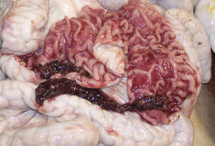
FIGURE 30-1 Gross lesions of the hemorrhagic form of proliferative ileitis.
(Courtesy Peter Moisan.)
Macroscopic lesions occur almost coincidentally in time with microscopic lesions. Microcolonies of L. intracellularis are found in apical cytoplasm of crypt epithelial cells during the first week postinfection, but enterocyte hyperplasia is not in evidence until nearly 1 week later and can continue for more than 6 weeks. Gross lesions are usually found in terminal ileum, but also in jejunum, cecum, and proximal colon. In the acute form, intestinal tissue is thickened and turgid, with a corrugated serosal surface and lumenal blood clots. Chronically affected intestines have irregular, patchy, subserosal edema, mainly at the mesenteric insertion. Ileal mucosa is thickened, with deep folds and patches of pseudomembrane. Surviving animals may have hypertrophic and thickened muscularis mucosa. Microscopic lesions consist of adenomatous proliferation of crypt enterocytes (Figure 30-2), in association with morphologically compatible organisms (Figure 30-3). Crypts are elongated and enlarged, and immature epithelial cells are highly mitotic. Goblet cells are reduced in numbers or absent in affected areas; inflammatory cell infiltration is minimal. Intestines of acutely affected pigs are congested, and blood is found in the intestinal lumen. Infected enterocytes usually have short irregular microvilli. Resolution of lesions is closely related to disappearance of intracellular organisms. Inflammation is a factor only in later-stage lesions and is not characteristic of the primary lesion. Following initiation of mucosal hyperplasia in hamsters, there is progressive replacement of mature villous columnar absorptive cells by undifferentiated crypt-type cells.
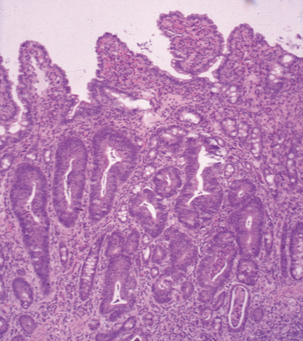
FIGURE 30-2 Proliferative lesions in the nonhemorhagic form of Lawsonia intracellularis infection.
(Courtesy Lynn A. Joens.)
Stay updated, free articles. Join our Telegram channel

Full access? Get Clinical Tree


