The Genus Leptospira

Chapter 31 The Genus Leptospira


Leptospirosis is encountered commonly by those dealing with animal infectious disease. Its occurrence as a zoonotic infection of humans is strongly influenced by connections to animal production, tropical climates, at-risk occupations, and behavior; it is counted by some as an emerging infectious disease of humans and by others as the most widespread zoonosis. The highest incidence of leptospirosis in the United States is in Hawaii. Interest in human leptospirosis has increased in recent times as a result of several large and well-publicized flood-associated case clusters in Central and South America. This might, as some have suggested, result from increased leptospiral virulence, but it is perhaps more likely to be caused by altered interaction among humans, reservoir species, and the environment.


Manifestations of human disease vary from nearly subclinical flulike illness to fulminant and fatal pulmonary hemorrhage and hepatic and renal failure. Icteric leptospirosis with renal failure was reported in the nineteenth century, and although much of the credit for this has been given to Adolf Weil, the description of the syndrome predates his work. Leptospirosis was recognized as an occupational disease in ancient China, and akiyami (autumn fever) is a part of modern Japanese medicine. Early in the twentieth century, physicians studying yellow fever in New Orleans found spirochetes in tissues of jaundiced patients and briefly advocated an etiologic role for them. The link between spirochetes and what we now know as leptospirosis was established later, when Japanese and German workers detected spirochetes and specific antibodies in the blood of miners and soldiers with infectious jaundice.


Leptospirosis is strongly environmentally associated, and transmission depends on direct or indirect interactions between humans and mammalian (usually rodent) reservoir hosts. Leptospira interrogans serovar icterohaemorrhagiae may have reached western Europe in the 1700s, coincident with extension of the range of Rattus norvegicus from Eurasia; the role of rats in epidemiology of the disease was discovered early in the twentieth century.


Epidemiologic patterns of leptospirosis fall roughly into three categories. Few serovars are involved in human disease in temperate climates, and infection is inevitably associated with direct contact with infected farm animals. In wet tropical areas, many more serovars infect humans, domestic animals, and large numbers of reservoir species; human exposure is often caused by widespread environmental contamination. Also, rodent-borne leptospirosis occurs in the urban environment, especially with infrastructure disruption by natural or man-made disasters. Human leptospirosis has not been a reportable disease in the United States for nearly 10 years, but urban disease in humans is not uncommon and is frequently misdiagnosed.


Until recently, the genus Leptospira was divided into species interrogans (the name deriving from the question mark shape of the organism and comprising pathogenic strains) and biflexa (encom-passing saprophytic, mainly environmental, strains). Growth at 13° C, 8-azaguanine resistance, and failure to form spherical cells in 1 M NaCl distinguished L. biflexa from L. interrogans. Both species were divided into numerous serovars (>60 in L. biflexa and >200 in L. interrogans), and related serovars are placed in serogroups. Many serovars appear to have a certain species as a natural host, but animals and humans can be infected with a wide variety of serovars. The serovars causing disease in animals vary among countries and sometimes among regions in the same country. Most infections in domestic animals are caused by only a few serovars.


Genetic typing of leptospirae has yielded 13 genomospecies (Table 31-1), and this is likely the future of leptospiral taxonomy. However, there is little correlation between serologic type and genomospecies; it is not uncommon for serogroups and even serovars to be represented in multiple genomospecies. Furthermore, methods for genomospeciation are not widely available, and it seems likely that the antigen-based approach to classification will be the standard for some time to come.


DISEASES, EPIDEMIOLOGY, AND PATHOGENESIS


A few key concepts form the foundation for understanding leptospirosis and its pathogenesis. First, leptospires often colonize proximal convoluted kidney tubules and may be excreted in urine for extended periods by reservoir hosts without clinical signs (Figure 31-1). The carrier state may be as short as a few days or may extend throughout the life of the animal. This is considered a major means of host-to-host transmission. Indirect exposure depends on environmental moisture, neutral soil pH, and a sufficiently mild climate to favor survival of leptospires. Streams and ponds contaminated by the urine of wild rodents or domestic and wild animals can be a source of infection for domestic animals and humans, as can urine aerosols in milking parlors (especially those of the herringbone configuration). The organism can also be isolated from milk of infected cows, and this probably serves as a means of transmission to humans and calves (Figure 31-2).
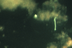


Second, localization in tissues from which the organism cannot be readily shed is common in infection of at least some hosts with specific serovars. In some urban areas more than 90% of Norway rats have serovar icterohaemorrhagiae localized in brain, and horses not infrequently experience localization in the eye.


Third, many serovars of leptospirae can be roughly categorized as host adapted or nonadapted. Infections by the former tend to be relatively mild and sporadic, with venereal transmission and lifelong colonization of the genitourinary tract; serovars hardjo in cattle and bratislava and tarassovi in swine are examples. Nonadapted strains, however, are more likely to produce catastrophic infections, with abortion storms in pregnant animals and, not infrequently, death of adult hosts. The carrier state is generally brief. Serovar pomona is nonadapted for swine and cattle, as is serovar canicola for dogs. Serovars of serogroups Icterohaemorrhagiae and Ballum are adapted to rats, the latter adapted to mice. The relationship between hosts and adapted strains gives rise to a minor but long-lasting serologic response, whereas nonadapted strains provoke high antibody titers. It should be obvious that a serovar may be adapted to one species and not others. It seems reasonable to speculate that most, if not all, species have host-adapted strains, including perhaps humans.


Fourth, certain steps seems to be common to Leptospira-induced disease, although all need not occur in every case and in some infections can lead only to very mild clinical signs. Exposure by mucosal or conjunctival routes, or through broken skin, is followed by invasion and eventual development of leptospiremia. Organisms proliferate in parenchymatous organs, including liver, kidneys, spleen, and meninges. Venereal transmission may result only in colonization of reproductive organs. The period of leptospiremia may be shorter (with host-adapted strains) or longer (with nonadapted strains), but in either case gives rise to circulating antibodies that mediate clearance of leptospirae by complement-mediated bacteriolysis. Damage to the maternal-fetal interface or active invasion beyond the placenta may cause fetal death and abortion, or birth of weak offspring. Fetuses infected in the third trimester (and perhaps earlier) may be born with specific antibodies. Organisms are cleared by the effects of antibodies and complement, or if antimicrobial therapy is applied, but may localize for days to years in kidney, reproductive tract, brain, or other tissues.


Fifth, mechanisms of pathogenesis are largely unknown, although the recently demonstrated ability to genetic transform leptospirae should facilitate detailed analysis of leptospiral virulence genes. Leptospiral endotoxin is of low potency, compared with that of many other gram-negative bacteria. Lipopolysaccharide (LPS) mediates adherence of neutrophils to endothelial cells and platelets, suggesting a role in development of thrombocytopenia. LPS is immunogenic and responsible for serovar specificity. In acute leptospirosis, damage to vascular endothelium is common, with resulting hemorrhage and disseminated intravascular coagulation (DIC). The last may be mediated, at least in part, by endotoxin, but sphingomyelinase hemolysins produced by many serovars may be responsible for this vascular damage and for specific clinical signs such as hemoglobinuria. Hemolysis by organisms of serovar lai is not related to sphingomyelinase activity, but is apparently due to a pore-forming protein. Protein and glycoprotein cytotoxins have been described.


Leptospiral attachment to epithelial cells can be enhanced by antibody. Virulent strains can bind to fibronectin. On the negative side (for the leptospirae), antibodies and complement are opsonizing; the outer envelope may be antiphagocytic. Leptospires induce apoptosis in vivo and in vitro.


Some symptoms may be immune mediated. Immune complexes may be associated with central nervous system (CNS) inflammation, although this apparently does not occur in the kidney. Autoantibodies, including IgG anticardiolipin antibodies, are detected in acute human illness. Antileptospiral antibodies cross-reacting with equine ocular tissues are apparently involved in pathogenesis of recurrent uveitis because retinal damage relates to the presence of B lymphocytes in the retina.


Finally, there is sufficient leptospiral promiscuity in host selection that it is difficult to say with certainty that a given serovar cannot infect a specific host. Certain strains are more commonly associated than others with disease in a given species, but even that varies with geographic region. Thus it is probably best for veterinary practitioners and diagnosticians to become familiar with the serovars infecting animals in the specific locale in which they practice (Table 31-2).


TABLE 31-2 Examples of Leptospira Serovars Associated with Disease in Specific Hosts


Serovar Hosts and Associated Disease
L. kennewicki Equine abortion, repeat breeding (United States, Northern Ireland, England)
L. bratislava Equine abortion, repeat breeding (United States, Northern Ireland, England)
Porcine abortion, infertility (worldwide)
L. pomona Porcine, bovine, equine abortion
Skunks
L. canicola Canine renal, systemic disease
Porcine systemic disease, abortion
L. icterohaemorrhagiae Canine septicemia
Bovine, porcine abortion
Classic association with rats
L. hardjo Bovine abortion, infertility
L. grippotyphosa Canine renal, systemic disease
Wildlife infections, especially raccoons, skunks


Disease in Cattle


Cattle are commonly infected by serovars hardjo and pomona, although infections by serovars grippotyphosa, icterohaemorrhagiae, and canicola are not uncommon. Serovar hardjo is apparently adapted to the bovine host, and produces sporadic abortions and infertility, often without other clinical signs. Fetal expulsion is delayed by several days after fetal death as a result of serovar hardjo infection. Colonization of the female upper reproductive tract is as common as that of kidneys. Thus the organism is available locally for interaction with embryos. Localization in the seminal vesicle is also quite common, and bulls and cows likely pass the infection back and forth at breeding. A common misconception is that transmission cannot occur under dry conditions. Given venereal transmission and the potential for exposure at congregation sites (such as water sources), hardjo and other serovars are quite efficiently transmitted in arid environments. Cows may experience mild to moderate mastitis during the acute phase of disease.


Infection by nonadapted strains often produces more dramatic signs. Infected animals are usually febrile, can be anemic and icteric, are often hemoglobinuric, and may be diarrheic. Abortion is common, with immediate expulsion of the dead fetus. Depending on the nature of the exposure, the number of animals in a group, and management of remaining animals after initial cases, abortions may occur because storms affect as much as 90% of pregnant females in a herd. With early recognition it is sometimes possible to interrupt the progress of an outbreak by administration of antimicrobials such as streptomycin, but in most cases they have no positive effect.

< div class='tao-gold-member'>

Stay updated, free articles. Join our Telegram channel

Jul 18, 2016 | Posted by in PHARMACOLOGY, TOXICOLOGY & THERAPEUTICS | Comments Off on The Genus Leptospira

Full access? Get Clinical Tree

Get Clinical Tree app for offline access