The Genera Escherichia and Shigella

Chapter 13 The Genera Escherichia and Shigella


The genera Escherichia and Shigella are class γ-proteobacteria, in the order Enterobacteriales and family Enterobacteriaceae.


THE GENUS ESCHERICHIA


The genus Escherichia was named in honor of German pediatrician Theodor Escherich, who first isolated the type species, Escherichia coli. Five additional species within the genus are Escherichia albertii, Escherichia blattae, Escherichia fergusonii, Escherichia hermannii, and Escherichia vulneris, but E. coli is the most significant pathogen. Escherichiae are straight gram-negative medium to long rods ranging from 0.4 to 0.7 μm by 1 to 3 μm, and occurring singly or in pairs. They have both oxidative and fermentative metabolism and are oxidase negative and catalase positive. Acid and gas are produced from D-glucose and most species are motile.


Diseases and Epidemiology


Escherichia coli inhabits the lower ileum and large intestine of most vertebrates, with colonization of the neonatal gastrointestinal tract occurring within hours of birth. At about 108 organisms per gram, it is the predominant facultatively anaerobic bacterium isolated from the feces. Survival and multiplication of E. coli in the environment makes “coliform” counts valuable indices of fecal contamination, especially in testing water for potability.


Most strains of E. coli are of low virulence and associated with opportunistic infections, whereas others are highly virulent. Different strains cause different types of disease, so it is important to discriminate between pathogenic and nonpathogenic strains. Serologic classification, based on seroreactivity of E. coli surface molecules, was developed for this purpose. It is especially useful for identifying strains that cause intestinal disease and has been used extensively for epidemiologic purposes. Two E. coli surface components that are the basis for serologic classification are the O antigen of the lipopolysaccharide and the H antigen of the flagellum. The O antigen determines the sero-group and the H antigen the serotype. A capsular antigen (K) may also be used for classification.


Escherichia coli are primary pathogens causing enteritis and septicemia in a variety of domestic species, including poultry, pigs, ruminants, dogs, cats, horses, and rabbits. Other important diseases are ruminant mastitis, canine pyometra and cystitis, and oomphalitis in young chicks. Infections may be endogenous or exogenous in nature. Extraintestinal disease is generally caused by the animal’s normal microbial flora, whereas strains causing gastroenteritis are usually from an exogenous source.


Strains of E. coli associated with disease in the gastrointestinal tract are classified based on virulence properties (Table 13-1). At least six different groups or pathotypes are recognized, including enterotoxigenic (ETEC), enteropathogenic (EPEC), enterohemorrhagic (EHEC), necrotoxigenic (NTEC), enteroinvasive (EIEC), and enteroaggregative (EAggEC). EAggEC and EIEC strains have not been reported from domestic animals. The former is associated with persistent diarrhea in humans in developing countries. Food- or water-borne outbreaks of human diarrhea have been attributed to EIEC.


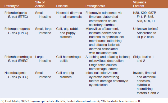
ETEC strains are generally species specific (Figure 13-1). They adhere via fimbriae and elaborate cytotoxins. In neonatal mammals they cause acute, watery diarrhea that may be followed by terminal bacteremia and remain an important cause of economic loss for cattle and swine producers. Clinical signs include white, yellow, or gray scours that occur during the first week of life, and microscopic examination of distal jejunum and proximal ileum reveals bacterial colonization of the mucosal epithelium, without villus atrophy. Some ETEC strains express K88 or F18 fimbriae, colonizing small intestine and producing diarrhea in recently weaned pigs. The common fimbrial adhesins and serogroups associated with disease are in Table 13-2.


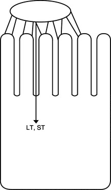
TABLE 13-2 Fimbrial Adhesins and Serogroups Associated with ETEC in Domestic Animals


FimbriaI Adhesin Animal Host Serogroups
K88 (F4) Pig O8, O141, O147, O149
K99 (F5) Pig, calf, lamb O8, O9, O20, O107
987P (F6) Pig O9, O20, O141
F41 Pig, calf O9, O101
F18 Pig O138, O139, O141
F1845 Calf O101
F17 Calf, lamb, kid

EPECs (Figure 13-2) are isolated from humans and farm animals, as well as dogs, cats, and rabbits, in all of which they produce attaching and effacing lesions. Strains are emerging as important causes of diarrhea in puppies and in other animals, and are associated with “failure to thrive” syndrome. Diarrhea is often mucoid and chronic, as opposed to the watery diarrhea associated with ETEC. Histologically, attaching-effacing lesions are the hallmark of disease.


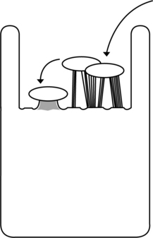
Many EHEC strains (Figure 13-3) produce Shiga toxin, which is similar to the cytotoxin of Shigella dysenteriae O1. These strains are often referred to as Shiga toxigenic E. coli(STEC) or verotoxigenic E. coli(VTEC), with the latter name derived from toxin effects on vero cells in culture. EHEC and STEC are important causes of diarrhea in calves less than 8 weeks of age. Affected animals exhibit mucoid to bloody diarrhea that is rarely fatal, but leaves calves dehydrated, weak, and stunted.


Edema disease, also known as “gut edema” or “bowel edema,” is a communicable enterotoxemia that results in significant death losses in recently weaned pigs. It is caused by EHEC-like bacteria that have acquired the genes for production of a Shiga toxin variant called Stx2e. Strains carrying the F18ab fimbrial type, originally described as F107, are most often associated with this condition. Unweaned pigs may acquire infection in the farrowing house, but lack of full expression of the F18 receptor in piglets younger than 20 days of age augurs against disease in neonates. Clinical signs include ataxia, paddling, confusion, palpebral edema, and sudden death. Postmortem examination reveals edema in colonic mesentery, submucosa of the glandular cardiac portion of the stomach, gallbladder and small intestine, and areas of cerebellar hemorrhage. Adherent gram-negative rods are seen in small intestine, and degenerative angiopathy in small arteries and arterioles, swelling of endothelial cells, and focal encephalomalacia in brainstem are common.


Escherichia coli O157:H7 is the most common EHEC serotype implicated in cases of human diarrhea and hemorrhagic colitis. Hemolytic-uremic syndrome (HUS) is a complication of EHEC infection, characterized by acquired nonimmune hemolytic anemia, thrombocytopenia, and acute renal failure. Infection with O157:H7 is zoonotic. As many as one third of domestic ruminants are asymptomatic carriers and represent the principal reservoir for human infection. Fecal shedding is transient, often lasting 1 to 3 months. Swine, horses, and deer can also be carriers of E. coli O157:H7, but do not appear to be major sources. Transmission occurs through consumption of undercooked meat, unpasteurized dairy products, and vegetables or water contaminated by feces of carrier animals. Epidemiologic investigations have clearly linked human infection to consumption of contaminated and improperly cooked beef. On occasion, O157:H7 has been associated with hemolytic-uremic syndrome in racing greyhounds fed undercooked or raw ground beef.


Cytotoxic necrotizing factors 1 and 2 are elaborated by NTEC. NTEC 1 strains have been recovered from humans, cattle, piglets, dogs, cats, and horses in association with diarrhea, urinary tract infection, and septicemia. NTEC 2 strains have been isolated from diarrheic and septicemic cattle and sheep.


Several fimbriated strains of E. coli are associated with intestinal, as well as extraintestinal disease. The F17 group of fimbriae has three epitopes. F17a fimbriae are isolated from bovine ETEC strains, septicemia in lambs and calves is associated with the F17b type, and F17c strains are recovered from septicemic and diarrheic cattle. The last are also associated with lamb nephrosis syndrome. F18 strains producing Shiga-like toxins cause swine edema disease.


Colibacillosis or colisepticemia of poultry is a common systemic disease with significant economic impact. Bacteremia follows intestinal infection or respiratory exposure to high numbers of organisms in the environment. The infection is characterized by acute septicemia or subacute airsacculitis and polyserositis in poults and young broilers. Adverse environmental conditions or concurrent infectious diseases predispose to clinical disease. Signs are nonspecific and age of affected birds, duration of illness, and affected organs vary from case to case. In 4- to 8-week-old birds, acute death may be preceded by anorexia and lethargy. Postmortem lesions include hepatitis and splenitis, with increased fluid accumulations in the thoracic and abdominal cavities. Survivors of acute septicemia usually develop pericarditis, perihepatitis, and fibrinopurulent airsacculitis, and the latter is the hallmark of colibacillosis. Other less common disease manifestations are pneumonia, salpingitis, arthritis, and osteomyelitis.


Peracute bovine mastitis most frequently results from a coliform infection, with E. coli being the most commonly recovered pathogen. Disease is characterized by rapid onset of fulminating infection, with painful swelling of the udder, production of milk with a serumlike consistency, lethargy, anorexia, and signs of endotoxemia (including fever, cold extremities, brick-colored mucous membranes, shock, and sudden death). Mastitis typically occurs during the early postpartum or peak production period and is reflective of heavy fecal contamination of the environment. Disease tends to be confined to cattle housed in restricted space. Predisposing factors may include anatomic abnormalities in the teat or udder, wet or dirty loafing areas, udder trauma, and unsanitary or malfunctioning milking equipment or techniques.


Escherichia coli is the organism recovered most frequently from the infected uterus of dogs and cats. Pyometra occurs more commonly in dogs than in cats. Affected bitches tend to be middle aged or older, whereas younger queens may develop disease. Canine and feline pyometra most often develops during the luteal phase of the cycle or after the administration of progestins. Examination of isolates recovered from cases of pyometra are similar to corresponding fecal isolates, suggesting that fecal contamination of the vaginal vault may be a predisposing factor. Clinical signs in bitches may include vaginal discharge, anorexia, lethargy, polyuria, polydipsia, and vomiting. Vaginal discharge and abdominal bloating are the most common signs in cats. Septicemia and endotoxemia may be sequelae in untreated animals.


The organism is also a common cause of canine cystitis. As with pyometra, affected animals are likely to harbor the same strain in their intestinal tracts, vagina, or prepuce, and infections are typically ascending. Predisposing factors include urolithiasis, anatomic or neurologic abnormalities of the urinary tract, neoplasia, indwelling urinary catheters, or prolonged antimicrobial therapy. Clinical signs include dysuria, polyuria, hematuria, and production of foul-smelling cloudy urine. In many instances cystitis is asymptomatic, manifesting simply as bacteriuria. Untreated disease can progress to pyelonephritis.


The spread of virulent strains of E. coli in the farm environment is presumed to occur through feed, aerosols, fomites, and carrier animals. Factors that predispose to disease include failed passive transfer in neonates, intensive husbandry practices, poor sanitation, and stressors such as dietary changes, cold weather, and commingling of animals from different sources. Neonates less than 1 week of age are especially prone to infection due to their lack of a fully developed immune response and established intestinal microflora. In addition, receptors for certain E. coli adhesins are present only for the first few days or weeks of life. Sudden dietary changes at weaning, including increased protein and decreased fiber levels, can result in a massive overgrowth of enterotoxigenic or Shiga toxin–producing strains. Inheritance of an autosomal recessive gene in large white breeds such as Yorkshire and Landrace is correlated with genetic predisposition to edema disease.

< div class='tao-gold-member'>

Stay updated, free articles. Join our Telegram channel

Jul 18, 2016 | Posted by in PHARMACOLOGY, TOXICOLOGY & THERAPEUTICS | Comments Off on The Genera Escherichia and Shigella

Full access? Get Clinical Tree

Get Clinical Tree app for offline access