Chapter 2 The operating room
Introduction
The operating room for extraocular surgeries is usually the standard operating room with more specialized illumination. Limited magnification, provided by head loupes and spectacle-mounted telescopes, is usually sufficient for surgeries of the orbit, eyelids, nasolacrimal system, nictitating membrane and conjunctiva. However, in the operating room where corneal and intraocular surgeries are routinely performed, in addition to the operating microscope, special instruments often include the phacoemulsification unit, ophthalmic cautery, cryotherapy, laser instrumentation, and retinal detachment surgery instrumentation.
Depending on the number and type of ophthalmic surgical procedures performed daily or weekly, the composition of the operating environment will vary. The operating microscope is the largest single investment, but will last for a very long time with proper care. To the full-time veterinary ophthalmologist, the operating microscope is indispensable for corneal and intraocular surgeries in all animal species.
A cabinet is maintained within the operating room for ophthalmic surgery, with special sterile instruments, individually wrapped, and ready for use. I prefer limited numbers of ophthalmic instruments arranged as external, minor, and major intraocular surgical packs. Another method divides the eye instrument packs based on intended use. Additional special instruments, individually wrapped and sterile, can be opened when needed; this reduces the wear and tear of cleaning and sterilization on instruments used infrequently but vital for certain eye surgeries. The external eye surgical pack is used to drape the surgical area, expose the globe, perform the lateral canthotomy, and appose the surgical wound. The external eye instrument pack should contain small towel clamps, a small saline bowl, a silicone irrigator for solutions to keep the cornea and conjunctiva moist, a few small hemostats, ophthalmic tissue forceps, strabismus, utility or tenotomy scissors for ocular tissues and sutures, small serrated and 1 × 2 teeth thumb forceps, knife handle and blades, and one or more eyelid specula.
The minor intraocular surgical pack provides the essential surgical instruments for corneal, glaucoma, and iris–ciliary body surgeries. The instruments in this pack can also be used to perform conjunctival grafts, superficial keratectomies, and primary closure of corneal ulcerations, and to treat partial to full-thickness corneal lacerations with an iris prolapse. This pack contains exclusively ophthalmic instruments, with small serrated, 1 × 2 teeth and tying forceps, curved and straight ocular scissors, cyclodialysis spatula, standard and micro-ophthalmic needle holder, corneal and anterior chamber irrigator, and portable battery-powered cautery unit. Special ophthalmic instruments, such as a corneal separator and iris scissors individually wrapped, sterile and ready for use, should be available within the operating room.
The major intraocular surgical packs provide the instrumentation for cataract and lens removal, and posterior segment diseases (mainly vitreous). The instruments in this pack include corneal section (cataract or corneoscleral) scissors, different types of tissue and tying forceps, the lens loop and spoon, cyclodialysis spatula, cautery unit, two or more cannulas, extracapsular lens forceps, forceps for tearing of the anterior lens capsule (capsulorhexis), and one or two different size needle holders. Other instruments, individually wrapped and sterile, should include iris scissors, intraocular scissors, and intraocular forceps. Some backup instruments should be available in case contamination or malfunction of instruments occurs during the surgical procedure.
chapter 1.2, chapter 1.3, chapter 1.4, chapter 1.5 and Box 1.1 list the different instruments for the varying ophthalmic surgeries. For more specialized surgeries, such as retinal detachment surgeries (see Table 1.5), instruments are often wrapped and sterilized separately.
Magnification
For nearly 40 years veterinary ophthalmic surgery has been progressively refined, and microsurgical procedures performed under the operating microscope, that started in the early 1980s, are now commonplace for surgery of the cornea and intraocular structures. Microsurgery for human ophthalmology started slowly in the 1950s by Perrit. The first commercial operating microscope was produced by Zeiss and featured coaxial illumination. Important refinements by Barraquer (XY mechanism) and Troutman (motorized zoom) defined the basic components of the modern operating microscope. The development of microsurgical instruments, sutures, and needles also stimulated the concurrent requirement for magnification. Surgical procedures of the extraocular tissues including the orbit, eyelids, nasolacrimal, and tear systems are still traditionally non-microsurgical; however, microsurgery involving the conjunctiva, cornea, and intraocular tissues has become common because of the small ophthalmic needles and sutures, and the need for exact apposition of the involved tissues. Initial use of the operating microscope may be somewhat frustrating and may prolong the surgical procedure. However, with patience and practice, the veterinarian will quickly appreciate the advantages of corneal and intraocular surgeries performed under magnification.
Head-mounted magnifiers
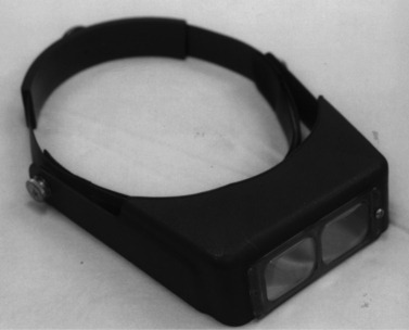
The exact requirements for magnification vary and are often influenced by the surgical patient load and the different types of ophthalmic surgical procedure performed. The simplest and least expensive magnification device is the binocular magnifier loupe worn on the head (Fig. 2.1). These head loupes can be used over prescription glasses. The head loupe is available in a number of different magnifications. Generally the lower magnifications are the most versatile because at the higher magnifications the focal length of the loupe is reduced, thereby limiting the working distance between the surgeon and the operating field. For instance, the head loupe with the 1.5× magnification has a focal length of 51 cm; the 1.75× magnification has a focal length of 35.5 cm; the 2× magnification is in focus at 25.5 cm; and the 2.5× magnification has a focal length of 20.5 cm.
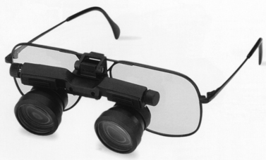
Individual telescope magnifiers are another low-cost alternative, and are generally recommended. They can be added to head loupes or attached directly to spectacles. These units permit accommodation for different interpupillary distances, allow use of prescription glasses, and can be elevated when not in use (Fig. 2.2). Although these units are lightweight, use for several hours when attached to spectacles can be very tiring.
Operating microscopes
Most veterinarians interested in corneal, intraocular, and vitreoretinal surgeries will eventually invest in an operating microscope. Once proficiency is achieved using the operating microscope, corneal and intraocular surgeries can be performed easily and quickly. The microsurgical ophthalmic instruments and very small 7-0 to 12-0 sutures can be easily manipulated with some magnification. The microscopic details provided during corneal incisions and wound apposition enhance the possibility of successful surgeries.
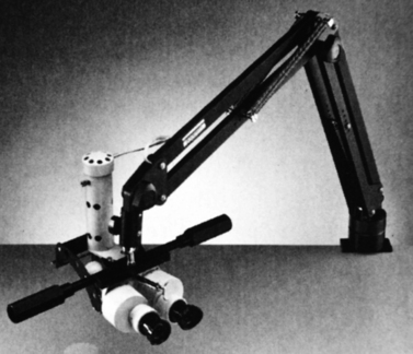
Operating microscopes can be portable and attached to either a table or floor base with casters. Table units are the least expensive, usually provide observation for only the surgeon, and changes in focus require manual adjustments (Fig. 2.3). Stationary ceiling-mounted operating microscopes are used infrequently in veterinary ophthalmology because of the need for portability between operating rooms. Most veterinarians use the floor-based operating microscopes which are very stable but mobile (Fig. 2.4).

Fig. 2.3 Portable table-mounted operating microscope. Unit weighs 9 kg and can be clamped to the surgery table. Working distance is 20 cm.

Fig. 2.4 Floor-based operating microscope. Unit provides for viewing by the surgeon, assistant surgeon, video recorder, TV and digital camera.
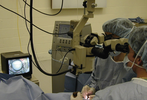
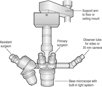
The operating microscope has several standard parts (Fig. 2.5). The base and mount are usually quite heavy and vary depending on whether the unit is table, floor or ceiling mounted. Various supporting arms permit adjustment of the operating microscope’s main body over the patient’s eye and angulation of the scope to the surgical field. With a large footplate that contains several switches, the surgeon can raise and lower the main body of the operating microscope to permit motorized coarse and fine focus of the surgical field. The scope’s main body consists of the focus and zoom systems, and a beam splitter that permits observation of the surgical field by the surgeon and assistant surgeon, and often a video recorder or 35 mm or digital camera. The fine focus of the surgical field and zoom or magnifying system are also controlled by a foot pedal. This allows slight adjustments on magnification and/or focus without interrupting surgery.
As with the head-mounted magnifiers, the magnification of the operating field is variable and inversely related to the focal length. The range of working distance between the patient’s eye and the base of most operating microscopes is 125–500 mm, with 200–250 mm the most common distance. The larger microscopes provide for dual observations for the surgeon and an assistant, and often an additional observer or camera (35 mm or digital SLR cameras or video recorder). Adjustments in magnification (zoom) and focal length are usually achieved by foot controls that change the focus up and down, and change the magnification (zoom). Magnifications with these operating microscopes vary, but generally range from 3× to 15× or 20×. Most scopes have built-in zoom systems that permit immediate changes in magnification during surgery. Other models have different fixed steps of magnifications that require manual changes. With the operating microscopes with different fixed magnifications, parfocalization is important to avoid marked variations in focus during changes in magnification.
To accommodate changes in both magnification and focus, the center of the surgical field should be in the middle of the microscope’s field, and the surgical area should be relatively level. Hence, the animal eye is carefully positioned so that the entire cornea, anterior chamber and iris surface are at the same levels of focus. Resolution of surgical field is optimal in the center of the scope’s optical system. Changes in the amount of magnification directly influence the size of the surgical field. With the 12.5× eyepiece, the 125 mm objective provides a surgical view of about 20 mm diameter; with the 200 mm objective the surgical view is 33 mm diameter; with the 300 mm objective, the surgical view is 50 mm diameter; and with the 500 mm objective, the surgical view is about 80 mm diameter. The most comfortable magnification and working distance is generally in the 175× range. Although higher magnifications may provide additional details, they reduce the working space as well as the depth of field.
The magnification also influences the depth of the surgical field; as magnification increases, the depth of the field decreases. During cataract surgery in the dog or cat, some change in the operating microscope focus should be anticipated. Initially at least one area of focus is the cornea and the incision into the anterior chamber, and later a second area of focus is the anterior lens for tearing and removal of the anterior lens capsule. The magnification may need to be changed further to permit visualization of the posterior lens capsule. I recommend the lower range of magnification for the surgeon initially which accommodates longer working distances between the operating microscope and the patient’s eye, and greater depth of the surgical field for most corneal and intraocular surgeries. This usually permits visualization of the entire eye and the palpebral fissure. With experience, the higher magnification or the zoom feature of the microscope can be used, but generally with the surgical field no smaller than the cornea. Some operating microscopes possess sterile caps for both the objectives (to permit small adjustments in the interpupillary distances and the focus of each objective) and the base of the microscope (however, as a general rule, if the surgical instrument touches anything except the patient’s eye and draped instrument table, it is discarded and a new instrument substituted).
Illumination systems are usually of two types: 1) the primary light system is incorporated into the operating microscope permitting direct illumination of the eye that is especially helpful during surgery in the posterior chamber and in the vitreous space; and 2) an ancillary light system mounted next to the operating microscope body that directs light to the eye at a slight angle. These light systems also function as a reserve; if one bulb malfunctions, the surgical field will continue to be illuminated. Both systems should have heat-absorbing filters to shield the eye as much as possible. Often the main and accessory light systems possess rheostats, permitting independent changes in the intensity of illumination. Retinal phototoxicity may occur with prolonged and intense illumination of the ocular fundus. The minimum level of illumination to adequately perform the surgery is the best guide.
Because of the magnification, positioning of the eye for surgery is important. With 10× to 20× magnification the depth of field is limited. As a result, the dog or cat is placed in dorsal recumbency, the head is stabilized by a U-shaped vacuum pillow or sandbags, and the operated eye is positioned in the center of the operating microscope’s field, with the cornea, anterior chamber, iris, and lens surfaces within focus. Other species, for instance the horse, are placed in lateral recumbency, and vacuum pillows or sandbags are used to position the head so that the eye is parallel to the operating microscope and the different areas of the cornea are in the same focus. If the eye is not level with the operating microscope, surgery in different places on the cornea or elsewhere will require intermittent changes in focus throughout the entire surgery. To simplify the optics of the operating microscope, maintaining both the scope and the patient in a vertical plane (rather than at an angle) will permit the majority, if not all, of the surgical field to remain in simultaneous focus.
Illumination
Adequate illumination is essential for ophthalmic surgery. The dark ocular tissues, especially the anterior uvea, require high intensity focal illumination for visibility. Traditional sources of light for ophthalmic surgery include overhead surgical lamps, primarily for extraocular surgical procedures; portable and ceiling-mounted ’cool lamps’ that filter the majority of infrared light waves that dry the tissues, used for both extraocular and intraocular surgical procedures; and direct and fiberoptic light systems that are routed through the operating microscope during microsurgery. Small focal operating and examining lamps that are head mounted may also be considered; however, because of their weight, they are generally used for only short periods of time. Many different models of each illumination system are available; choice of the surgical lamp is also influenced by the anticipated frequency of use and costs. As a general rule, once the ocular tissues are illuminated during ophthalmic surgery, intermittent irrigation of the corneal and conjunctival surfaces with sterile 0.9% saline or balanced salt solution should be performed to prevent corneal and conjunctival epithelial damage.
Chairs for microsurgery
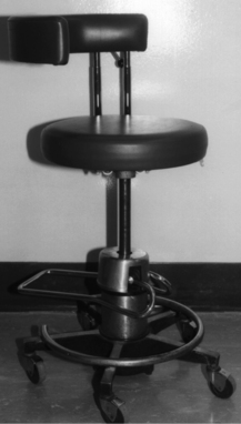
All corneal and intraocular surgical procedures in small animals are performed with the surgeon and assistant surgeon seated on adjustable stools with casters (Fig. 2.6). The height of the stools should be adjustable to accommodate different surgeons as well as the height of the operating table and the patient. The recommended operating room chairs can be adjusted with a hydraulic activated foot pedal, permitting changes during surgery without interrupting surgery. In some stools the back rest can be rotated 180° and moved to the front of the surgeon to provide arm rests during microsurgery. These arm rests can be covered with sterile stockinet. The operating chairs for the surgeon and assistant surgeon should be comfortable and help to avoid fatigue that can adversely impact surgery.
Patient preparation
Preparation of the skin of the eyelids and conjunctiva differs from that of most other general surgical procedures. During cleansing and preparation of the eyelid skin, these agents often contact the cornea and conjunctiva. As a result, solutions such as iodine in alcohol (Lugol’s solution) and isopropyl alcohol routinely used for skin preparation in general surgery are avoided as they are very toxic to the corneal and conjunctival epithelia, and cause immediate epithelial sloughing.
Clipping of hair and eye protection
The hair and eyelashes are carefully clipped with small electric hair clippers. The eyelashes may be coated with petroleum jelly or ophthalmic ointment, and carefully clipped by small sharp scissors. While removing the hair of the eyelids and adjacent skin about the orbit, one should avoid creating any nicks and abrasions as these wounds can cause unnecessary irritation and unsightly swellings postoperatively.
Several different strategies have evolved to protect the eye during surgical preparation of the eyelids and/or conjunctiva. Liberal amounts of ophthalmic ointment or petroleum jelly can be applied to the cornea just before clipping the eyelid hair. Hairs that fall onto the cornea and conjunctiva become embedded in the petroleum jelly, which is carefully removed later with sterile cotton swabs. A reusable plastic contact lens coated with ointment can also be used for this purpose. The contact lens is removed immediately before surgery along with any hair and debris from surgical preparation.
Cleansing and disinfection
The eyelids and adjacent orbital skin are cleaned and prepared for corneal and intraocular surgery. A mild antiseptic solution containing aqueous 0.5% povidone–iodine is the recommended ocular surface disinfectant for all ophthalmic surgical procedures. Chlorhexidine diacetate (0.05% and 0.5%) is toxic to the canine eye and causes chemosis, corneal epithelial edema, and corneal erosions. However, chlorhexidine gluconate (0.05%) with 4% isopropyl alcohol is both a safe and effective antimicrobial disinfectant for the dog’s cornea and conjunctiva.
Both povidone–iodine and chlorhexidine are bactericidal. The 0.5% aqueous dilution of povidone–iodine produces rapid broad-spectrum antimicrobial effects against the commonly isolated Staphylococcus aureus, Staphylococcus epidermidis, α-hemolytic Streptococcus sp., and Escherichia coli, as well as many fungi and viruses in the dog. At effective antimicrobial dilutions, such as the 0.5% level, povidone–iodine does not cause corneal epithelial edema, corneal epithelial sloughing, eyelid edema, or conjunctival irritation in the dog. After at least two 1-minute cleansing periods with povidone–iodine, the eyelids, conjunctiva, and cornea are liberally flushed with sterile 0.9% saline or balanced salt solution. Sterile cotton-tipped swabs are used to remove any remaining exudates and hair from the conjunctival fornices and surfaces, and the ophthalmic surgical site is ready for draping.
The 5% povidone surgical scrub, which contains 4.5% alcohol, provides an excellent germicidal preparation of the facial skin (as for parotid duct transposition), but cannot safely be used for the eyelids. This preparation is toxic to the corneal epithelium, producing a generalized loss of this layer (essentially a chemical superficial keratectomy).
Most corneal and intraocular surgeries are performed with the pre-existing ophthalmic condition requiring medication within the operating room. Accordingly, the eye is often medicated immediately preoperatively, during the operative procedure, and following surgery. Often the preoperative condition or the immediate postoperative inflammation can be substantially reduced as a result of this perioperative medication of the eye and adjacent structures. Recent studies in dogs indicate that bacterial contamination of the anterior chamber occurs in about 30% of dogs undergoing cataract surgery, and topical as well as systemic antibiotics should be administered. Antibiotic therapy for full-thickness corneal perforations and lacerations is often administered during the surgical correction in the solutions used to irrigate the external ocular surfaces, and for re-establishment of the anterior chamber after repair of the corneal defect. Fortunately, bacterial endophthalmitis is very rare in the dog after intraocular surgery.
Head positioning
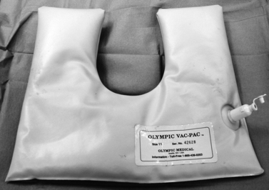
The majority of corneal and intraocular surgical procedures are performed with the small animal patient in dorsal recumbency. If head-mounted magnifiers are worn, the dog may be positioned in lateral recumbency. Ropes and adhesive tape are used to position the animal’s legs. Although the duration of most ophthalmic surgical procedures is less than 1 h, a circulating water heating pad between the patient and the surgery table reduces the possibility of hypothermia. It is important to place the animal’s head in a secure position to prevent any positional changes during surgery. Towels, water bottles, or several small low-cost sandbags can be used to maintain the head in the desired position. An alternative superior scheme uses a vacuum bead-filled U-shaped pack for stabilizing the head (Fig. 2.7). The patient’s head is positioned on the vacuum pack, and the pack is manipulated to provide the selected head position. Once the proper position is achieved, vacuum is applied temporarily to the pack. Once the air is removed, the pack becomes very rigid, holding the head in a fixed position. Release of the vacuum postoperatively causes the pack to return to a soft and moldable structure.
Draping
Once surgical preparation of the eye and associated structures is complete, the area is carefully draped for surgery. Simultaneous use of both paper (barrier) and reusable cloth (to absorb any moisture) drapes is recommended. Plastic drapes with self-adherent backings and rubber dental dam material can also be employed immediately about the surgical site. Cloth drapes are used to absorb the irrigation solutions, but unfortunately allow contamination with the other non-sterile surfaces of both patient and operating table. Paper or barrier drapes repel all moisture and prevent contamination from the other areas, and are most often used.
Our standard draping procedure is to position four small surgical towels about the surgical site to absorb the irrigation solutions, and secure them with small bulldog towel clamps. An additional large paper drape with a small hole to provide adequate surgical exposure is placed over these towels and secured with additional towel clamps. As with any surgical procedure, draping is designed to provide adequate exposure of only the operative site and a barrier to the adjacent non-sterile areas.
Irrigation
To prevent drying of the conjunctival and corneal surfaces during ophthalmic surgery, small volumes of sterile 0.9% saline, balanced salt solution, lactated Ringer’s solution, or other physiologic solutions are used intermittently to moisten the ocular tissues. Both the pH and tonicity of these solutions should approximate the tear and aqueous humor fluids. Inadequate levels of moisture provided during eye surgery will result in unnecessary corneal epithelial damage and possible limitations on the use of certain classes of medications postoperatively.
Sponges/swabs
To assist with hemostasis and removal of material during ophthalmic surgery, small sterile cotton swabs are used routinely for corneal surgeries, and for the extraocular aspects of intraocular surgeries. For use inside the anterior chamber during intraocular procedures and full-thickness corneal surgical procedures, disposable cellulose sponges (often triangular shaped) are recommended. Cotton swabs are avoided during intraocular surgical procedures because some of the small cotton fibers may loosen and enter the anterior chamber, increasing the intensity of postoperative anterior uveitis.
Basic operative approach
For corneal and intraocular surgeries, exposure of the entire cornea and anterior segment is preferred. In most small animals under general anesthesia, the globe rotates downward and inward. This may be advantageous for corneal and anterior segment surgeries involving the dorsal and dorsolateral regions, but not when access and visibility of the entire cornea and anterior segment are necessary. With less than adequate surgical exposure, the duration of surgery is prolonged and the procedure may be more difficult to perform.
Retrobulbar injections in dogs
Access to the cornea and anterior globe may present exposure problems in small animals, especially in certain breeds of dog. Fortunately, the lateral and dorsolateral aspects of the dog orbit are incomplete, and accommodate retrobulbar injections. Injections of sterile 0.9% saline can enhance the presentation of the cornea and globe, but only with some risk.
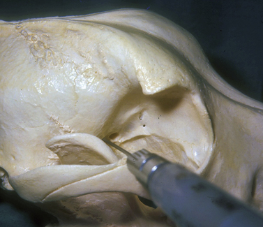
The injection is performed with the dog under general anesthesia with the objective of forcing the globe further rostrad or forward in the orbit, or to turn the globe and improve exposure of a selected area of the cornea and/or anterior segment. The amount of sterile saline injected is ascertained as the injection is performed and the response of the globe to the space-occupying solution. The hypodermic needle may be inserted caudal to the junction of the lateral orbital ligament and dorsal aspects of the zygomatic arch (Fig. 2.8). The needle is directed towards the retrobulbar space in a ventromedial direction toward the opposite mandibular joint. The solution may be injected in the lateral aspects of the extraocular muscle cone, or immediately caudal to the globe and within the retrobulbar muscle mass. Injections external to the retrobulbar muscle cone will rotate the globe laterally; injections immediately behind the globe will push the globe forward. The volume injected should be limited to produce the desired outcome but not result in undue pressure and distortion of the globe.

Fig. 2.8 An 8 cm, 22 g hypodermic needle is inserted dorsal to the zygomatic arch and caudal to the lateral orbital ligament, and directed toward the opposite mandibular joint in the dog. A variable volume of sterile saline can be injected in the animal under general anesthesia to force the globe forward.
Another injection site is ventral to the anterior zygomatic arch and rostrad to the vertical portion of the ramus of the mandible (see Chapter 3). The hypodermic needle, after passing the ramus of the mandible, is directed toward the orbital fissure. Injections external to the retrobulbar muscle cone in the orbital floor and the medial orbit wall are possible with this method, and can be used to shift the globe dorsally.
Retrobulbar injections in cats
Retrobulbar injections in the cat are not recommended because of the limited retrobulbar space and difficulty in proper positioning of the injection.
Retrobulbar injections in horses
Retrobulbar local anesthetic injections have been described in the horse by Berge and Lichenstern. The posterior orbit and entry of the critical cranial nerves in the horse is about as deep as in cattle, but the posterior orbit is more conical. With gas inhalation general anesthesia and often neuromuscular blocking agents and forced ventilation, retrobulbar nerve blocks in the horse are infrequent.
In the Berge method, an 8–10 cm, 18 g needle is inserted caudal to the supraorbital process of the frontal bone near the supraorbital foramen. The long needle is directed ventromedial (about 40° from the vertical) and slightly caudal toward the area of the orbital fissure where 15–20 mL of local anesthetic is injected (see Chapter 3).
In Lichenstern’s method, an 8–10 cm, 18 g needle is inserted 1.5 cm caudal to the middle of the supraorbital process. The needle is directed toward the opposite last upper premolar tooth. The taut extraocular muscles’ fascial cone may be felt as the needle penetrates it. Approximately 20 mL of local anesthetic is injected near the orbital fissure (see Chapter 3).
Of the large animal species, intraocular surgery is performed most often in the horse. As this species has considerable scleral elasticity (low scleral rigidity), sizeable retrobulbar injections can markedly indent the posterior segment and increase the likelihood of vitreous prolapse during cataract surgery.

Stay updated, free articles. Join our Telegram channel

Full access? Get Clinical Tree

