CHAPTER 24 The Male Reproductive Tract: Prostate, Testes, and Semen
THE PROSTATE GLAND
Collecting and Preparing Samples
Enlargement of the prostate gland is the primary reason for obtaining samples from it. Clinical signs suggesting that the prostate may be enlarged include difficult defecation or micturition, although the latter occurs less frequently. Sometimes urine is tinged red from blood, or blood or pus may drip from the penis. When either of these signs occurs, rectal palpation of the prostate may reveal symmetric or unilateral enlargement, focal bumpiness, or softness.1,2 Such prostate abnormalities detected by palpation or, occasionally, radiographic examination are the primary indications for obtaining material from the gland for cytologic evaluation.
Material from the prostate can be obtained directly through the urethra or by fine-needle aspiration of the gland. A direct method is to digitally massage the prostate while aspirating through a urinary catheter passed to the level of the gland.3,4 More prostatic material can be obtained by washing the part of the urethra near the prostate with a small amount of saline through gentle injection and aspiration while gently massaging the prostate. Material may also be obtained from the prostate by ejaculation.2,4 The prostate is gently massaged during the process to increase the amount of material of prostatic origin because this method may contain contaminants from other parts of the reproductive tract. The first part of the ejaculate contains material primarily derived from the prostate gland.2,4 The sample size is usually large and has a moderate concentration of cells. In addition to prostatic epithelial cells, ejaculated samples may contain numerous spermatozoa and cells of other reproductive tract and urethral structures. Simultaneously collecting and evaluating urethral wash specimens and urine samples (by antepubic cystocentesis) can alleviate some of the problems of interpreting the cytologic and microbiologic findings of the material collected by ejaculation.
The prostate can be sampled by direct aspiration with a small-gauge needle. Ultrasound-guided aspiration is superior to all other methods for obtaining specimens, especially from focal lesions such as cysts. The patient is sedated and restrained in dorsal recumbency in a padded, V-shaped trough. The inguinal area is anesthetized and the area is prepared as for surgery. Sterile petroleum jelly is applied to the skin to provide an acoustic coupler between the skin and the scanhead, and a small stab incision is made in the skin to facilitate needle passage. While the ultrasound scanhead is held stationary and needle advancement is monitored on the screen, the needle is directed to the prostate or a specific location within the prostate. When the needle has reached the proper site, a small amount of specimen is aspirated into a sterile syringe.4
The prostate can be sampled by digital guidance of a needle as well, which may be passed through the posterior abdominal wall or the perineal region, although the latter method has not been widely used in veterinary medicine. At either site, local anesthesia is necessary to prevent discomfort and facilitate needle guidance. When the prostate is so enlarged that it can be palpated through the abdominal wall, the needle can be guided to the prostate while the dog is in either lateral or dorsal recumbency and the prostate is immobilized with one hand. This method is usually effective only when the prostate is markedly enlarged.4 From the perineal area, a finger is placed in the rectum, and the needle is passed through the skin in the perineal area and guided into the prostate along the finger. With both methods, the gland is gently aspirated while the needle is moved within it.4 Occasionally, only a small amount of material is obtained by aspiration, but it is usually adequate for making one or two direct smears.
Normal Prostate
The cytologic features of the normal prostate vary depending upon the method of obtaining materials. Samples obtained by direct aspiration contain fewer contaminating cells and are usually more cellular than samples obtained from ejaculation, massage, or urethral washes. Glandular cells of the normal prostate are found in small to medium clusters and have centric nuclei and a finely stippled or reticular pattern. The cytoplasm stains slightly acidophilic and has a fine, granular appearance due to the very fine, basophilic material coursing through the cytoplasm (Figures 24-1 and 24-2).
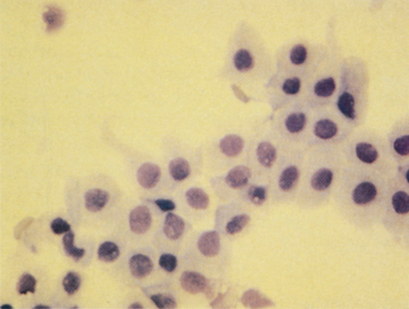
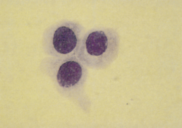
Cells and contaminating material from other locations in the urogenital tracts may be found.
Spermatozoa
Sperm heads characteristically stain blue-green with Wright’s method. Spermatozoa, which often adhere to other cells, possibly with many attached to a single epithelial cell, are most frequently found in ejaculated material, but can also be found in massage or wash samples (Figure 24-3).
Urothelial Cells
Urothelial cells (i.e., transitional epithelial cells) usually appear individually but may also be in small clusters. They are larger than prostatic epithelial cells and have homogenous, lightly basophilic cytoplasm and a lower nuclear-to-cytoplasmic ratio than prostatic cells (see Figure 23-1). Urothelial cells originate from the bladder and the tubular structures of the urinary and genital tracts and are most frequently found in samples obtained by ejaculation, massage, and washing.
Other Epithelial Cells
Cells of the ductus deferens and the epididymis are difficult to distinguish from prostatic cells.
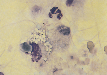
Ultrasound-Contact Gel
The gel used to ensure adequate acoustic coupling of the ultrasound scanhead to the body wall may contaminate the material obtained with ultrasound assistance. Small, azurophilic needle-like structures are found individually or in large clusters (Figure 24-4). When there is a large amount of material, the cells may be obscured or not be stained sufficiently for evaluation. The amount of contamination can be minimized by wiping excess gel from the skin before introducing the needle. Enough gel usually remains on the skin and scanhead to ensure sufficient contact for visualization of the prostate and accurate guidance of the needle.
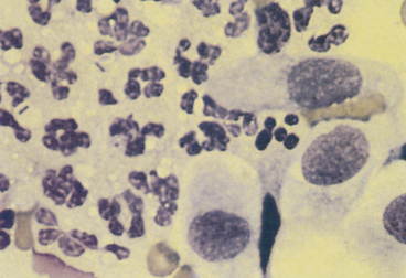
Prostatitis
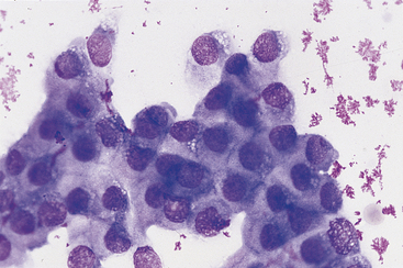
Purulent inflammation in which neutrophils dominate is the most frequent inflammatory lesion of the prostate. In septic prostatitis, the neutrophils have features, including karyolysis and foamy cytoplasm, that are suggestive of toxic degeneration. There usually are variable numbers of macrophages that typically have abundant, foamy cytoplasm. Bacteria may be found both intracellularly and extracellularly, and when they are found, it is necessary to determine whether they are actually the cause of the inflammation or are contaminants from other locations in the genital or urinary tract.3 If the sample was obtained by an aspiration technique, the bacteria should be considered the cause, but if the sample was obtained through the urogenital ductal system, it is necessary to determine if the bacteria are contaminants from the external genitalia or from an inflammatory lesion elsewhere in the urinary or genital system. Intracellular bacteria are certainly a strong indication that the inflammatory process is septic. In addition to inflammatory cells, clusters of various sizes of epithelial cells may be obtained. In aspirates obtained from inflamed prostates, prostatic cells appear to have loose cohesion. Additionally, their cytoplasm usually shows increased basophilia, indicating that the prostatic epithelium is hyperplastic secondary to the inflammation (Figure 24-5).
Stay updated, free articles. Join our Telegram channel

Full access? Get Clinical Tree


