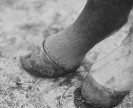
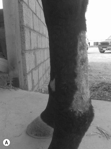
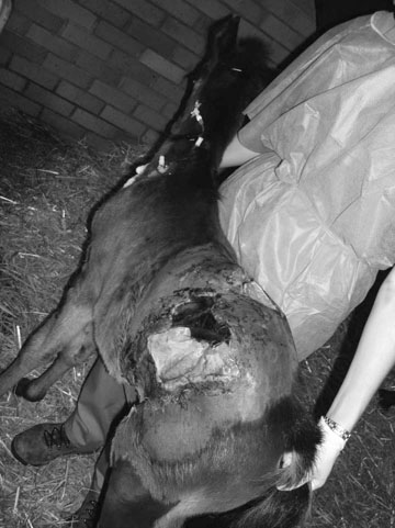
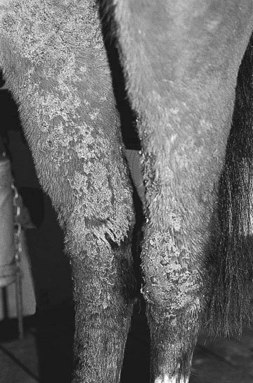
• Clinical signs are diagnostic (there are no other disorders with these signs evident at birth). • If the affected area is small treatment with repeated surgery may have a satisfactory result. However, most cases involve extensive lesions with resulting secondary infections and septicemia a common problem. • Euthanasia is frequently recommended if the condition involves large areas. • DNA testing is available and will also differentiate carriers. • Affected horses have an elevation of their urine deoxypyridinoline (DPD):pyridinoline (PYD) ratios which is diagnostic of the disease, from birth, but this testing may not be readily available. • Currently, there is no effective therapy for HERDA. Affected horses are often euthanized due to the severity of lesions and associated discomfort. • Selenium poisoning (affects mane and tail initially and is frequently accompanied by sloughing of hooves) • Chronic anhidrosis (history of lack/reduced sweating) • Ectoparasites (pruritis results in damage and loss of hair especially of mane and tail) • Zinc, copper and iodine deficiencies (rare causes of hair loss; body hair is affected before the mane or tail) • Alopecia areata (mostly affects the skin of the body and head) • Diagnosis is usually made based on clinical signs and breed, but histopathology of biopsy samples may help to demonstrate the lack of functional hair follicles. • It is important to differentiate from selenium poisoning. • No treatment is possible and it does not affect the horse clinically, being more of a cosmetic defect. • Systemic lupus erythematous-like syndrome (pigmentary incontinence and multisystemic involvement) • Vitiligo (loss of pigmentation on the face and perineum) • Appaloosa parentage syndrome (appaloosa cross-bred foals frequently do not have color change until 3–4 years of age, at which time a fading in coat color and development of white spots can be seen) • Leukoderma/leukotrichia following trauma or radiation therapy • Discoid lupus (photo-exacerbated condition with discoid epidermal lesions on the face and limbs). • Clinical signs are usually diagnostic, but radiography or ultrasonography can be used to confirm. • Biopsy shows multinodular deposits of minerals within fibrous and granulomatous tissue. • Complete surgical removal is the treatment of choice, but depending on the area affected and the size of the lesion may be difficult to achieve. • Nodular panniculitis/steatitis (painful inflammatory changes in the panniculus or subcutaneous fat; commonly affects the girth and lower chest walls) • Clinical signs are suggestive but definitive diagnosis requires biopsy (excisional biopsy is required). • Eosinophilic granulomatous inflammation is detected histologically. • Surgical excision is curative but diagnosis should be definitive prior to excision. In many cases no treatment is pursued due to the benign nature of the condition. Healing of limb wounds is often slow, particularly if the injury, such as deep wire cuts, involves deeper tissues. Where the periosteal blood supply is disrupted sequestration of the underlying bone frequently occurs (see Fig. 7.121). Failure of even relatively minor wounds to heal at sites subjected to trauma involving (even minor) periosteal damage should be investigated carefully in case such sequestration has occurred. These wounds invariably completely fail to heal until either the sequestrum has been spontaneously reabsorbed or surgically removed. Other reasons for the failure of wounds to heal include excessive movement at the site, inadequate wound contraction, infection (bacterial and parasitic), poor or interrupted blood supply, foreign bodies, excessive blood loss and underlying systemic disease. Wounds in some areas such as the thorax and neck often heal rapidly, relying on a combination of wound contracture and effective spontaneous drainage of exudates and inflammatory debris. Wounds involving the eyelids (see p. 358) or the margins of the mouth, nose and vulva, may heal effectively by second intention healing but may result in a possibly more serious secondary effect after cicatrization (scarring) has taken place. • Zinc (Fig. 9.19). Zinc has a major influence on skin health and adult daily requirements are 500 mg. Deficiency causes a generalized alopecia and flaking of the superficial skin layers giving a dandruff-like appearance. Dietary supplementation with zinc methionine or other zinc salts is curative over a number of weeks. • Iodine. Young foals born to mares with sub-optimal iodine diets during pregnancy may be born with an obvious goiter (see p. 221) and such foals are often weak and have a very poor coat quality. On occasion they may be almost hairless. Iodine poisoning (iodism) results from ingestion of abnormal amounts of iodine (usually from supplements). Cutaneous findings are heavy surf/seborrhea mostly on the mane and tail base but it can also be seen on the body. Diagnosis is normally based on a history of iodine supplementation and treatment involves removal of the supplement. • Copper. Copper deficiency is particularly unusual in horses, even in areas with a primary copper-deficient soil or in areas with high soil molybdenum concentrations, but in the event that the animal is unable to maintain its copper status, alteration in the color of the hair (hypochromotrichia) is usually all that is seen. Dark-pigmented hair may become obviously russet in color. Other more serious consequences of copper deficiency are a tendency to arterial rupture and chronic anemia. Diagnosis can be made by blood and liver analysis of copper levels but frequently response to supplementation is used. It is important to remember that not all forms of copper are bioavailable and if deficiency is suspected consultation with an equine nutritionist is valuable in selecting or developing supplementation. • Selenium poisoning (Fig. 9.20). Selenium causes toxicity either in metallic base form (usually following over-enthusiastic feed supplementation) or from plants which either concentrate selenium or have increased selenium levels as a result of seleniferous soils. Loss of mane and tail hair occurs, as well as the prominent signs of laminitis. In some cases the laminitis may be severe enough to result in separation of the coronary band and sloughing of the hoof. Long-term toxicity or repeated episodes may result in obvious laminitic deformities of the hoof walls with converging growth lines. • Diagnosis is usually based on history. Skin biopsy changes and changes in the hoof wall are non-specific. • This condition is treated by removing the source of selenium combined with a dietary adjustment to include high-protein supplement. • The overall prognosis is guarded with full recoveries difficult to achieve.
The integumentary system
Congenital/developmental disorders
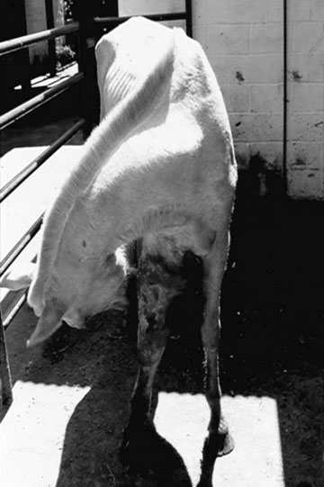
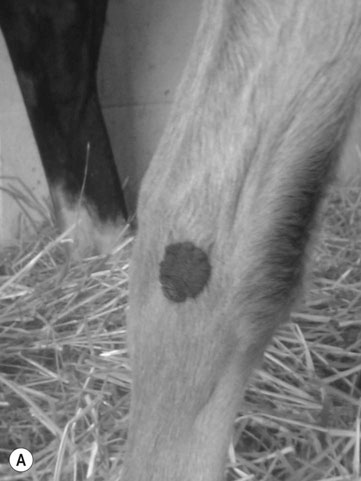
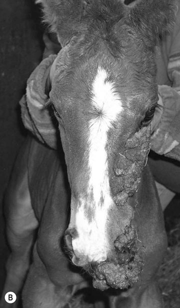
Epitheliogenesis imperfecta (Figs. 9.1 & 9.2)
Differential diagnosis:
Diagnosis and treatment
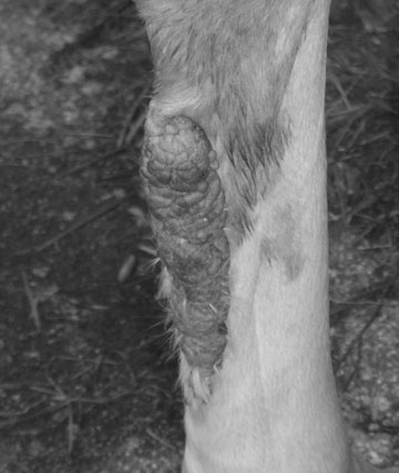
Congenital papillomatosis (neonatal wart/epidermal nevus) (Fig. 9.5)
Linear keratosis/alopecia (Figs. 9.6 & 9.7)
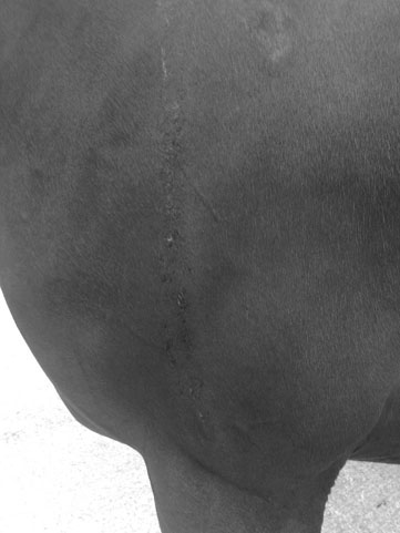
Differential diagnosis:
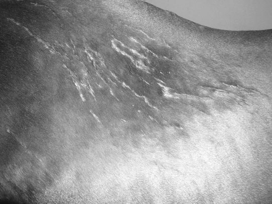
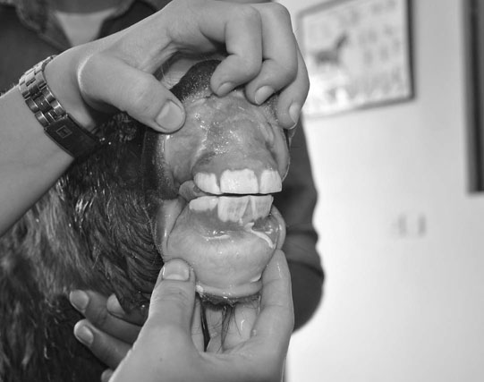
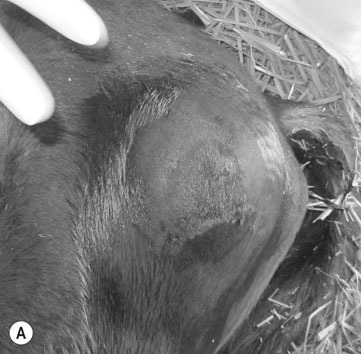
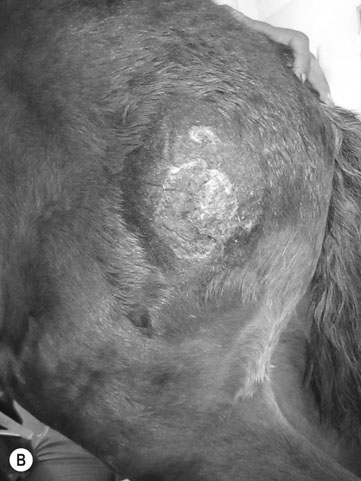
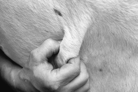
Hereditary equine regional dermal asthenia (HERDA; hypoelastosis cutis; cutaneous asthenia/Ehlers-Danlos syndrome) (Figs. 9.8 & 9.9)
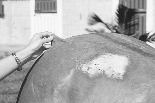
Differential diagnosis:
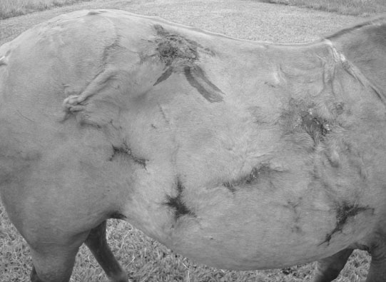
Diagnosis and treatment
Hypotrichosis/mane and tail dystrophy (follicular dysplasia) (Fig. 9.11)
Differential diagnosis:
Diagnosis and treatment
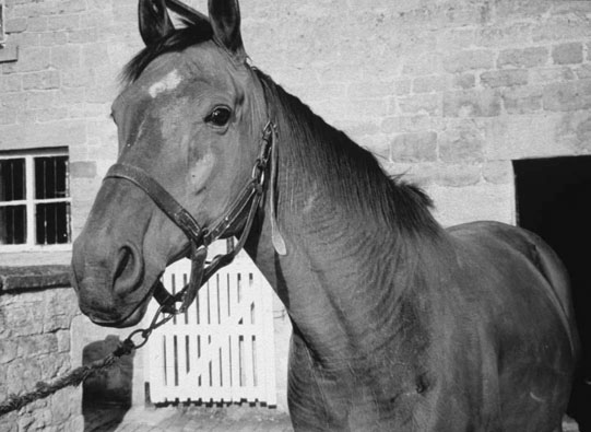
Arabian fading syndrome (pinky Arab syndrome) (Fig. 9.12)
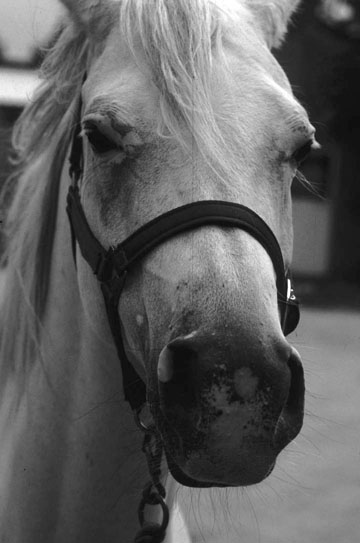
Differential diagnosis:
Non-infectious disorders
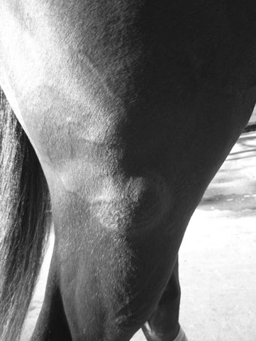
Calcinosis circumscripta (Figs. 9.13 & 9.14)
Differential diagnosis:
Diagnosis and treatment
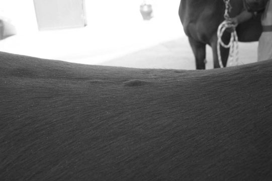
Equine axillary nodular necrosis (Fig. 9.15)
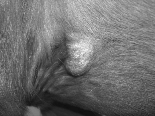
Differential diagnosis:
Diagnosis and treatment
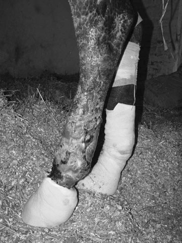
Wounds
Nutritional diseases with cutaneous manifestations
Diagnosis, treatment and prognosis of selenium poisoning
The integumentary system