Disorders of metabolism, nutrition and endocrine diseases


Disorders of metabolism, nutrition and endocrine diseases


Developmental disorders


Hypocalcemia


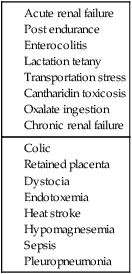
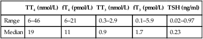
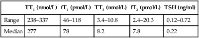
Calcium deficiency in the horse can be acute or chronic and result from a variety of conditions (Table 6.1). Chronic deficiency presents as abnormal cartilage and bone development with or without lameness.


Acute deficiencies result in clinical signs associated with neuromuscular excitability.


The clinical consequences that are seen are varied and will be discussed individually.


Retained placenta


While placental retention can occur for a number of reasons in some cases it may be confounded by hypocalcemia which results in decreased uterine tone and contractility. This has been further supported by the fact that mares treated with a combination of oxytocin and calcium borogluconate solution had a better response to therapy than mares treated with oxytocin alone.


Diagnosis and treatment of hypocalcemia


• The calculated deficit should ideally be based on the ionized calcium concentration but most laboratories measure total calcium.


• When treating hypocalcemia it is important to consider:


• Many horses with ionized hypocalcemia may only have mild undectable signs but may progress to develop other signs such as ileus.


• Horses with functional kidneys can eliminate large amounts of calcium rapidly and it is rare to induce hypercalcemia from calcium administration but rapid administration could potentially result in cardiovascular complications. Current recommendations for speed of replacement are 1–2 mg/kg/h.


Hypercalcemic disorders
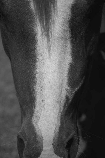
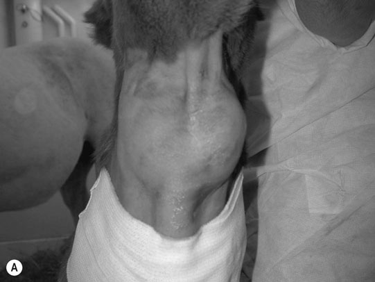
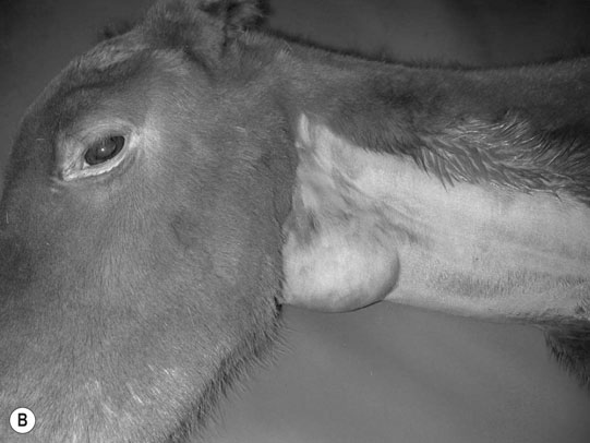
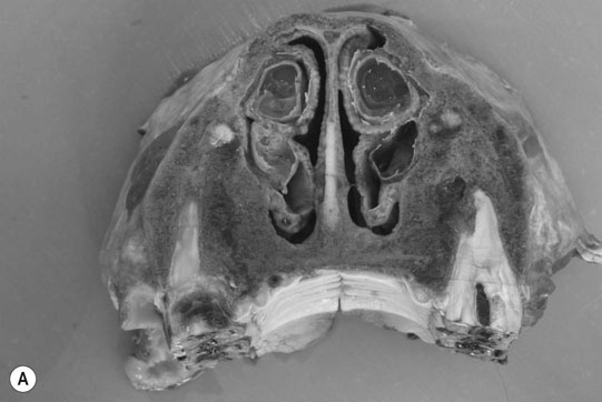
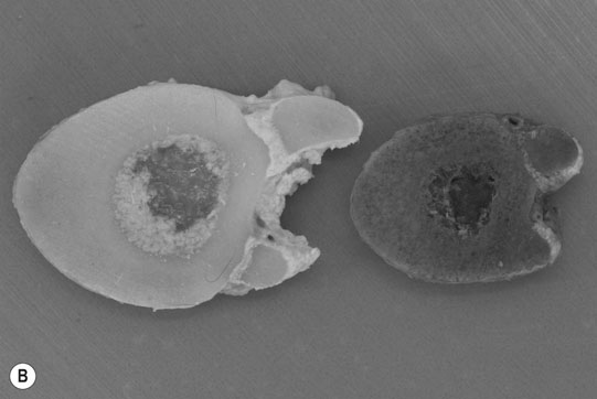
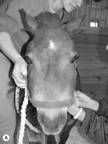
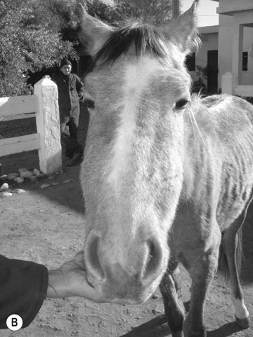
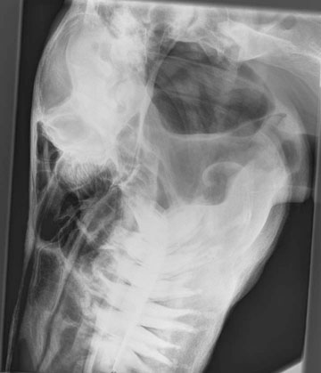
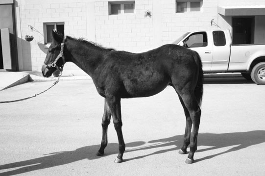

Primary hyperparathyroidism (Figs. 6.16.4)


An abnormality of the parathyroid gland results in excessive synthesis and secretion of PTH with no response to negative feedback from Ca2+. Theoretically, adenoma or hyperplasia of the parathyroid gland could result in this condition but histological examination of the gland is often complicated by the fact that it is difficult to find due to its small size and variable location.


image
Figure 6.3 Radiographs of a forelimb (same horse as Figs. 6.1 and 6.2). Note loss of radiodensity. The mare later fractured a cannon bone and was subjected to euthanasia.

image
image
Figure 6.4 (A, B) Necropsy (same horse as Figs. 6.16.3). Note changes of osteodystrophy fibrosa involving limbs and head. (B) Bone from normal pony on left and affected pony on the right.

The condition has been reported in association with osteodystrophia fibrosa. These horses have enlarged facial bones, poor body condition and lameness.


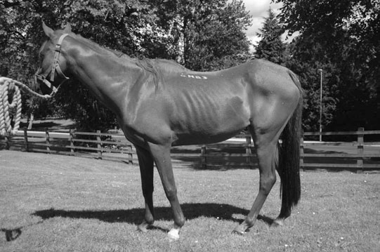
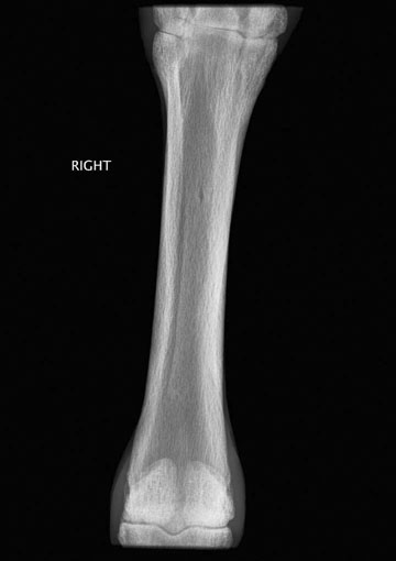
Secondary hyperparathyroidism (Figs. 6.56.7)


Excessive secretion of PTH occurs in response to hypocalcemia, hyperphosphatemia or hypovitaminosis D from renal dysfunction or nutritional imbalances. Renal secondary hyperparathyroidism does not appear to be important or common in horses, perhaps due to the fact that most horses with renal failure are actually hypercalcemic rather than hypocalemic in other species.


Nutritional secondary hyperparathyroidism (bran disease, big-head, Miller’s disease)

Horses fed a diet low in calcium, high in phosphorus or both may develop this condition.


Traditionally this disorder was associated with excessive grain feeding but due to improvements in nutrition is not as frequently encountered in developed countries. However, ingestion of substances which bind out the available calcium, such as phytic acid in wheat-bran or oxalate in tropical pasture grasses in Australia and North America, render the calcium less available and results in a further relative oversupply of phosphate. The most obvious manifestation of the disease is the development of a marked thickening of the flat bones of the head due to the more or less symmetrical laying down of cartilage in place of bone. Although young growing horses are most severely affected older horses are also subject to the condition. Shifting-leg lameness due to tendon and ligament avulsions, micro-fractures or even more serious pathological fractures are commonly the first sign of the disease. The extent of cartilage deposition and the consequent weakness of the alveolar attachments of the molar teeth is a serious effect of the disease. Ultimately, cachexia develops, which is primarily a consequence of pain and physical obstructions to mastication.


Iron deficiency


Primary deficiency as a result of low dietary intake or poor absorption is rare but chronic blood loss, chronic infections or internal neoplasia often result in the secondary development of a dramatic, progressive and typical (microcytic, hypochromic) iron-deficiency anemia. In these horses the total body iron is invariably normal but the availability is severely impaired by the complex of inflammatory processes. Horses which suffer from a prolonged microcytic, hypochromic anemia without evidence of bone marrow activity should be investigated thoroughly for a possible underlying infectious or neoplastic disorder.


Thyroid disease


Thyroid gland function can be assessed by measurement of serum concentrations of thyroid hormones, either at rest or in response to thyrotropin-releasing hormone (TRH) or thyroid-stimulating hormone (TSH). Stimulation tests are regarded as more reliable but have the disadvantages of being more time-consuming and there is limited availability of TRH or TSH in an affordable form.


Bound or free forms of T3 or T4 can be measured in readily available assays (see Tables 6.2 and 6.3). It is best to measure levels of free hormone as changes in protein binding can alter the total level.


Certain drugs (phenylbutazone, corticosteroids), physiological events (strenuous exercise, diets high in energy, protein, zinc or copper) and pathological events (inappetance, non-thyroid disease) can alter the circulating levels of thyroid hormones without the presence of disease in the gland itself.


Hyperthyroidism


Hyperthyroidism is an extremely rare disorder in horses and the cases reported in the literature were attributed to thyroid gland neoplasias. Clinical signs in these cases included weight loss, tachycardia, hyperactive behavior, ravenous appetite and cachexia. Exposure to iodine, such as in topical blisters, can result in a temporary increase in circulating thyroid hormones but these cases are not associated with clinical signs of disease given above.


Hypothyroidism (Figs. 6.86.10)


Primary hypothyroidism in the horse is probably much less common than previously thought. Many of the clinical signs attributed to hypothyroidism such as obesity, abnormal fat deposits, subfertility and recurrent laminitis have now been recognized as part of the equine metabolic syndrome and many of the horses that were previously diagnosed as hypothyroid are now acknowledged to be insulin-resistant. Further supporting this is the fact that cases reported to have hypothyroidism as a result of thyroid neoplasia have predominantly presented with changes in skin/haircoat and poor exercise tolerance. Additionally, thyroidectomy in young horses results in delayed physeal closure, decreased growth rate, cold intolerance, low rectal temperature, lethargy and coarse haircoat as well as other non-specific changes in serum phosphorus, cholesterol and triglyceride concentrations.


Stay updated, free articles. Join our Telegram channel

Feb 27, 2017 | Posted by in EQUINE MEDICINE | Comments Off on Disorders of metabolism, nutrition and endocrine diseases

Full access? Get Clinical Tree

Get Clinical Tree app for offline access