The Genus Clostridium

Chapter 33 The Genus Clostridium


Clostridia are large, sporulating, gram-positive, oxygen-tolerant to strictly anaerobic rods. Clostridium piliforme is the exception, in that it is gram negative. The source of clostridia can be exogenous (most commonly the soil) or endogenous (often from the intestinal tract). Most pathogenic clostridia produce one or more toxins, and direct or indirect evidence links many of these with pathogenesis (Table 33-1). Myonecrosis often follows mechanical injury, with clostridial spores germinating in damaged, ischemic muscle. Toxemia can also ensue when spores are deposited in wounds, or when spore-containing tissues are damaged. Disruption of normal flora by antibiotic therapy or sudden dietary changes may precede enteric infection, which is frequently accompanied by intestinal lesions; in some types of enterotoxemias, intestinal lesions are minimal. Disease can also result from ingestion of preformed clostridial toxins. Death is a frequent endpoint, and is often acute or peracute. Vaccination with bacterin-toxoids or toxoids is an effective prophylactic measure for many clostridial diseases.


TABLE 33-1 Animal Pathogenic Clostridia


Pathotype Species Disease
Neurotoxic C. botulinum Botulism
C. tetani Tetanus
Histotoxic C. perfringens Myonecrosis, gas gangrene
C. septicum Malignant edema, braxy
C. chauvoei Blackleg
C. novyi Myonecrosis, infectious necrotic hepatitis (black disease), bacillary hemoglobinuria (redwater)
C. sordellii Myonecrosis, enteritis
Enteric C. perfringens Enteritis, enterotoxemia
C. difficile Diarrhea, antibiotic-associated diarrhea, pseudomembranous colitis, colitis X
C. colinum Quail disease
C. spiroforme ı-Enterotoxemia
C. piliforme Tyzzer’s disease

Animal pathogenic clostridia can be conveniently categorized as neurotoxic, enteric, and histotoxic (see Table 33-1). Some (e.g., Clostridium perfringens) are heterogeneous and thus rather cosmopolitan in terms of hosts and systems affected, whereas others (e.g., Clostridium colinum) cause a single, well-defined syndrome.


NEUROTOXIC CLOSTRIDIA


Diseases and Epidemiology


Tetanus usually results from inoculation of a traumatic wound with spores of Clostridium tetani. Minor penetrating wounds or abrasions, surgical incisions, docking, castration or eartag wounds, injection sites, and postpartum lesions in the reproductive tract may be portals of entry. The wound may be trivial, even unnoticed, but the reduced oxygen and Eh resulting from necrosis are usually required. The typical source of C. tetani is soil, and both vegetative cells and spores are detected in the digestive tracts of animals. Disease occurs frequently in horses, less often in other herbivores, and infrequently in pigs and carnivores. Cattle may develop tetanus following growth of C. tetani in the rumen.


After germination in the wound, C. tetani produces a neurotoxin that causes the majority of the symptoms of tetanus. The incubation period ranges from 24 hours to 2 weeks, varying with the toxogenicity of the infecting strain, the rate at which toxin is transferred to target tissues, and host sensitivity. Ascending tetanus follows retrograde, intraaxonal transport of toxin along the peripheral motor nerves to the central nervous system (CNS). Toxin crosses the synapse and binds to presynaptic axonal terminals, causing motoneuron hyperactivity, with sustained spasms inthe innervated muscles. Other muscle groups are affected when toxin travels within the spinal cord. Descending tetanus results from vascular dissemination of toxin, and clinical effects often begin in sites distant from the infection.


Muscular tremor and increased stimulus response appear early in the course of the disease, and are followed by impaired muscle function in the head and neck. Difficulty in chewing and swallowing is sometimes exacerbated by trismus. In horses with tetanus, nostrils may be flared, the third eyelid retracted, and the ears stiffly erect. Permanent rigidity follows the tetanic spasms, and muscles of the back and tail become rigid, with orthotonus. Mechanical respiratory failure and death occur in a few days to 2 weeks.


Botulism is typically an intoxication with one of several serologically distinct neurotoxins that cause neuroparalysis by blocking acetylcholine release from cholinergic nerve endings. Sporesof Clostridium botulinum are found in soil and sediment, and on plants growing in contaminated soil. The primary form of botulism in humans has shifted from intoxication to toxicoinfection, primarily in the form of infant botulism (“floppy baby syndrome”). Toxicoinfectious botulism can arise from a variety of foods, but one of the most common is honey. Botulinum spores are a microbiologic bonus for bees collecting pollen and nectar, and even the low concentration of spores (approximately 0.25 spore/g) can lead to initiation of disease in infants. Toxicoinfectious botulism also occurs in users of illicit injected drugs, particularly so-called black tar heroin, especially when injection is intramuscular (“muscle popping”) rather than intravenous.


Clostridium botulinum producing toxin sero-types A and B are found in soil, and types C, D, E, F, and G are common in wet environments. Spores germinate in animal carcasses or rotting vegetation and produce enough toxin to cause disease outbreaks in ruminants, horses, mink, and fowl; other carnivores, swine, and fish are occasionally affected. The most common types areC and D, although this varies geographically and among animal species. Loin disease and lamziekte (cattle), limberneck and western duck sickness (waterfowl), and spinal typhus and shaker foal syndrome (horses) are distinctive names applied worldwide to the various clinical syndromes.


Phosphorus-deficient animals may develop pica and ingest botulinum toxin with the bones of animals. The common name lamziekte (lame sickness) is applied because lameness often accompanies phosphorus deficiency. Poultry litter can also be a source of toxin. The lethal dose of toxin orally is larger than that by other routes, probably because of degradation by rumenbacteria. Equine botulism is also most commonly associated with adulterated feed, but shaker foal syndrome follows toxin production in the gut of the affected animal.


Outbreaks of type C botulism in waterfowl may originate with, and be sustained by, toxin in tissues of dead invertebrates. Invertebrate larvae ingest toxin from vertebrate carcasses and are consumed by fowl. Sporadic outbreaks of type E botulism in wild birds may be associated with consumption of toxin-bearing fish. Dabbling ducks are commonly affected, as are shorebirds. Mortality in migratory populations can exceed 50,000 birds in a single season.


Type C botulism is the most common form in domestic poultry, and multiplication of C. botulinum in the gut may be responsible for some outbreaks. Botulism in farmed mink is also caused most often by type C.


Type D botulism occurs in cattle, and a 1990 outbreak on the Darling Downs in Queensland, Australia, killed nearly 20% of a herd of 30,000 cattle. Spent poultry litter, fed as a source of nonprotein nitrogen, was contaminated with dead birds. In this outbreak, deaths continued after contaminated feed had been removed from the ration, suggesting toxicoinfectious botulism due to multiplication of the organism in the rumen.


The gene for botulinum toxin resides on a bacteriophage in some strains of C. botulinum, and the finding of Clostridium baratii and Clostridium butyricum toxins, which are serologically and pharmacologically similar to botulinum toxin, suggests gene transfer via this mobile element.


Clinically, botulism is characterized by anorexia, incoordination, ataxia, and flaccid paralysis. Paralysis of the tongue and pharynx leads to difficulty in swallowing, and death eventually results from respiratory paralysis. Horses experience tremors, and paralysis of facial muscles is common in cattle. In waterfowl, loss ofability to fly and a drooping head (“limberneck”) are common. Chickens often have diarrhea.


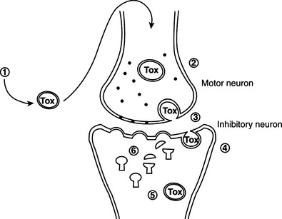
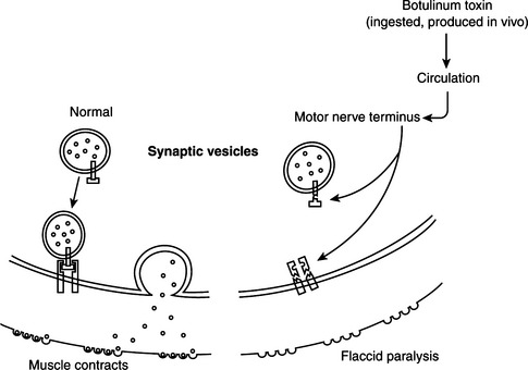
Pathogenesis of tetanus and botulism.


Neurotoxins of C. tetani and C. botulinum inhibit neurotransmitter release at CNS and peripheral synapses, respectively. Tetanus toxin bound to the presynaptic membrane of the neuromuscular junction is internalized and transported retroaxonally to the spinal cord. The result is spastic paralysis through toxin action on spinal inhibitory interneurons. The neurotoxins produced by C. botulinum are the most potent toxins known. They induce flaccid paralysis through intoxication of the neuromuscular junction.


These neurotoxins are zinc metalloproteinases that enter the cytoplasm and cleave specific protein components of the neuroexocytosis apparatus. They are composed of two disulfide-linked polypeptide chains, the larger of which is responsible for neurospecific binding and cell penetration. Upon reduction, the smaller chain is released into the cytoplasm, where its zinc-endopeptidase activity is expressed.


Tetanus neurotoxin and botulinum neurotoxins serotypes B, D, F, and G cleave VAMP/synaptobrevin, a membrane protein of the synaptic vesicles (Figures 33-1 and 33-2). Cleavage is atdifferent, single sites for each toxin. Botulinum A and E neurotoxins cleave synaptosomal-associated protein–25 (SNAP-25), a component of the presynaptic membrane, at two different carboxyl-terminal peptide bonds. Serotype C specifically cleaves syntaxin, another protein of the nerve plasmalemma. These three proteins are conserved from yeast to humans and are essential in a variety of docking and fusion events in every cell. The end result of toxin action on target cells is blockage of acetylcholine release, and muscle paralysis.


The target specificity of these toxins is based on interaction with the cleavage site and with a noncontiguous segment containing a structural motif common to VAMP, SNAP-25, and syntaxin. VAMP contains two copies of a nine-residue motif, also present in SNAP-25 and syntaxin. Antibodies against this motif cross-react among the three proteins, and inhibit the proteolytic activity of the neurotoxins.


Two additional C. botulinum toxins, designated C2and C3, are not neurotoxic. They are also binary toxins, but have ADP–ribosylating activity, similar to ι-toxins of C. perfringens and Clostridium spiroforme. In addition, they are much less toxic (mouse lethal dose values >45 ng) than botulinum toxin (minimum lethal dose [MLD] <0.01 ng).


HISTOTOXIC CLOSTRIDIA


Diseases, Epidemiology, and Pathogenesis


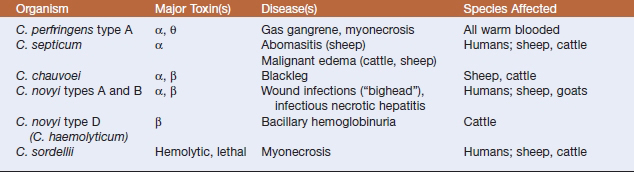
Histotoxic clostridia are common pathogens of humans and domestic animals, and a limited group causes most of the infections (Table 33-2). The hallmark is enthusiastic toxinogenesis, but common themes are acquisition of the infecting organism from soil or an endogenous source (such as the intestinal tract), entry to tissue following trauma, local multiplication and toxin production, occurrence of extensive local (and often systemic) tissue damage, and rapid death of the host. In animals, control by vaccination has decreased the incidence, and perhaps also the visibility, of some clostridial diseases, but renewed interest in mechanisms of pathogenesis, and to some degree the continuing interest inprevention of battlefield clostridial infections or intoxications, has yielded new information, particularly about modes of toxin action.


Clostridium septicum.


Clostridium septicum is commonly found in soil, and has been isolated from the feces of domestic animals and humans. It is frequently a postmortem invader from the gut, particularly in ruminants. The organism has been isolated from snails that play a role in the life cycle of liver flukes, as well as from flukes recovered from sheep experimentally infected with the parasites. Further, there is evidence that C. septicum can enter animals from the environment in one of the life stages of the fluke.


The organism causes malignant edema in humans, with infections often associated with traumatic wounds, occult bowel carcinomas, diabetes mellitus, liver cirrhosis, and peripheral vascular disease. Nontraumatic clostridial myonecrosis is an uncommon but often fatal condition that requires immediate medical and surgical therapy. Clinical features include rapidly evolving acute illness with pain, tachycardia, and bulla formation, followed by hypotension and acute renal failure; palpation and radiography reveal gas in soft tissues. Hemorrhage, edema, and necrosis develop rapidly as the infection spreads along muscular fascial planes. Early lesions are initially painful and warm, with pitting edema, but with time, the tissue becomes crepitant and cold. Death follows, often in less than 24 hours. The source of the organism is gastrointestinal in more than 50%of patients and the mortality rate varies from 33% to 58%, depending on method of management.


In addition to its role in myonecrosis, C. septicum may also cause enteric infections. In lambs or older sheep the organism penetrates theabomasal lining and produces a disease knownas braxy or bradsot, which is characterized byhemorrhagic, necrotic abomasitis and fatal bacteremia. Braxy causes heavy mortality in sheep in Great Britain, Ireland, Norway, Iceland, and the Faroe Island archipelago, and has been reported in Europe, Australia, the United States, and elsewhere. A similar disease syndrome occurs in calves.


Clostridium septicum infection can manifest as gangrenous dermatitis in chickens. Iatrogenic infections are more common in horses than in other species.


Malignant edema in domestic animals(Figure 33-3) usually follows direct contamination of a traumatic wound. Genital infectionscan be associated with mismanaged attempts at delivery, and umbilical infections are not infrequent in lambs. Hemorrhage, edema, and necrosis spread rapidly along fascial planes from the point of infection. As in human infections, the developing lesion is initially painful, warm, and it pits on pressure, but gradually becomes crepitant and cold, with loss of feeling. Death follows a period of fever, anorexia, and depression, often in less than 24 hours.
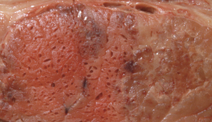


The pathogenetic mechanism by which C. septicum invades the abomasal lining (leading to development of braxy) is not known, but ingestion of cold or frozen feed is frequently an associated factor in both sheep and dairy calves; impaired mucosal function may allow entry of the organism, followed by local multiplication and dissemination throughout the body, producing local lesions and toxemia. Abomasal and proximal small intestinal walls are edematous, hemorrhagic, and sometimes necrotic.


Toxic or potentially toxic products of C. septicum include α-toxin (oxygen-stable hemolysin), β-toxin (DNase, leukocidin), γ-toxin (hyaluronidase),δ-toxin (oxygen-labile hemolysin), neuraminidase, chitinase, and sialidase. Recent work has demonstrated the pivotal role of α-toxin in pathogenesis of malignant edema and related infections. The effect of the purified toxin mimics some of the features of the animal and human diseases caused by C. septicum. α-Toxin is secreted as a 46 kD protoxin, which is activated by proteolytic cleavage and release of a 45 amino-acid fragment from the COOH terminus. The deduced primary sequence of α-toxin has 72% similarity (over a 387-residue region) with the primary structure of the Aeromonas hydrophila toxin aerolysin. There is good evidence that the propeptide is a chaperone, which stabilizes α-toxin monomers and escorts them to the membrane, where protease activation and oligomerization are followed by pore formation in the plasma membrane and colloidal-osmotic lysis. In vitro activation can be by trypsin cleavage, but furin and other eukaryotic proteases are involved in activation of toxin on the cell surface in vivo. α-Toxin likely binds to glycosylphosphatidylinositol-anchored protein receptors. Activated toxin forms 230 kD aggregates in erythrocyte membranes, causing release of potassium ions and hemoglobin.


A role for potential virulence attributes other than α-toxin has not been proven, but they may, in combination, increase capillary permeability and contribute to myonecrosis and systemic toxicity.


Clostridium novyi.


Clostridium novyi type Cis nontoxigenic (and therefore avirulent), but types A and B, as well as D (also called Clostridium haemolyticum), cause disease in humans and domestic animals. Differential production of α- and β-toxins determines toxin phenotype. However, C. novyi types B, C, and D may be one independent species arising from a single phylogenetic origin. 16S rDNA sequences of type B and D strains are identical, and are nearly identical to sequences from types C and A.


Strains of type A cause gas gangrene in humans and wound infections in animals. The hallmark lesion is edema, and “bighead” of young rams is illustrative; rapidly spreading edema of the head, neck, and cranial thorax follows bacterial invasion of subcutaneous tissues damaged by fighting. Clostridium novyi type A has been recently recognized with alarming frequency asa cause of septicemia in drug addicts who inject themselves intramuscularly.


Infectious necrotic hepatitis (“black disease”) of sheep and cattle results from C. novyi type B infection. Dormant spores germinate in liver tissue, often damaged by fluke migration, andsystemic effects with acute or peracute death follow dissemination of α-toxin. Its cardio-, neuro-, histo-, and hepatotoxic effects apparently produce edema, serosal effusion, and focal hepatic necrosis. The name “black disease” derives from the characteristic darkening of the underside of the skin as a result of venous congestion.


Clostridium novyi type D (C. haemolyticum) causes bacillary hemoglobinuria of cattle and other ruminants. Strains of type D resemble those of type B, except that type D strains produce no α-toxin and much more β-toxin.


Bacillary hemoglobinuria is most common in well-nourished animals more than 1 year of age. Deposition of type D spores or vegetative cells in the digestive tract and liver follows ingestion. Immature flukes migrate through the liver, causing hepatic necrosis and hypoxia, and inducing germination of spores in Kupffer cells. β-Toxin causes hepatic necrosis, and dissemination through the bloodstream leads to intravascular hemolysis and hemorrhage. Fever, pale mucous membranes, anorexia, abdominal pain, and hemoglobinuria (from which the common name “redwater” is derived) are typical clinical signs; when hemoglobinuria appears, 40% to 50% of red cells have been lysed, and death ultimately results from anoxia. Serosal effusions and a large circumscribed liver infarct are pathognomonic, and gram-positive rods are abundant in the sinusoids. Thus the pathogenesis of bacillary hemoglobinuria is similar to that of black disease of sheep, except that the primary toxin is β- rather than α-toxin. A typical case fatality rate is 90%to 95%.


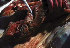
Type B may be involved in an emerging problem with sudden death in sows (Figure 33-4), which is associated with multiplication of the organism in the liver at or about the time of parturition. This disease has not been reproduced by experimental inoculation of sows.
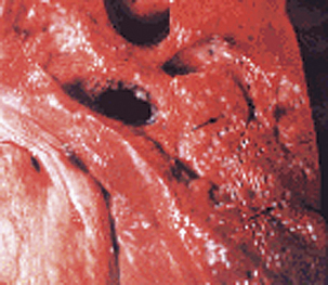


Clostridium novyi α-toxin causes rounding of cultured cells by effects on the cytoskeleton. It belongs to the family of large clostridial cytotoxins that modify small GTP-binding proteins. The substrate range includes N-acetylglucosamine and the active site is near the NH2-terminus. Full enzyme activity of the intact toxin resides on an approximately 550 amino-acid fragment, and mutation of aspartic acid residues within this fragment dramatically reduces enzyme activity. α-Toxin specifically modifies the Rho subfamily proteins Rho, Rac, Cdc42, and RhoG, by N-acetylglucosaminylation of a threonine molecule at position 37.


Little is known about pathogenesis of type D infections, but β-toxin probably plays a major role. It is a phospholipase C with lethal, necrotizing, and hemolytic activities. The phage that mediatesα-toxin production in type B can transducetype D to α-toxin production.


Clostridium chauvoei.


Blackleg is a necrotizing emphysematous myositis caused by Clostridium chauvoei. The organism inhabits primarily the intestines of cattle and sheep, although persistence in soil may be important, based on observed year-to-year occurrence of the disease on the same pastures. Clostridium chauvoei and C. septicum are closely related, based on sequence analysis of 16S rRNA genes.


Blackleg (Figure 33-5) occurs most commonly in well-fed cattle less than 3 years of age. Lesions usually occur in hindlimb muscle mass, but may be seen only in myocardium, or possibly the diaphragm or tongue. In sheep the diseasemore frequently presents as a wound infection resembling malignant edema (C. septicum infection) or gas gangrene (C. perfringens infection). Clinical signs include high fever, anorexia, depression, and lameness. Many lesions are internal, but superficial ones may be crepitant on palpation. Sudden death, without observed clinical signs, is common.


The periphery of lesions is typically edematous and hemorrhagic, with myonecrosis, whereas the central areas of the lesions are often dry and emphysematous. Bacterial production of butyric acid lends a rancid-butter odor to the lesions. Microscopically, evidence of leukocytic infiltration is negligible, but degenerative changes occur in muscle fibers, and edema, emphysema, and hemorrhage are common.


Ingestion is the most probable route ofexposure in cattle, and various tissues, especially skeletal muscle, are seeded with spores. Outbreaks may be due to spread of infection or a common source of local muscle anoxia, such as overexercise induced by encounters with phlebotomous insects. Damage to the muscle provides conditions favoring germination of dormant spores, with subsequent multiplication and toxin production. Acute indigestion may also initiate these events.


The roles of α-toxin, which is necrotizing, hemolytic, and lethal, and β-toxin, a DNase, remain undefined. Hyaluronidase (γ-toxin), oxygen-labile hemolysin (δ-toxin), and neuraminidase are of uncertain importance. Phase variation in motility and flagellation occurs, and flagellar expression is associated with virulence. fliC has been cloned and characterized, but immunization with recombinant flagellin protein does not protect mice against challenge.


Clostridium sordellii.


Clostridium sordellii isa common inhabitant of soil and of the intestine of domestic animals. Isolates have been obtained from myositis, liver disease, and sudden death in cattle, sheep, and horses. Edema of subcutaneous tissues and fascial planes, with subendocardial hemorrhage, is a common lesion, with terminal septicemia in experimentally infected cattle. Clostridium sordellii is sometimes found in the intestines of cattle experiencing “sudden death syndrome,” and bovine enteritis has been produced experimentally with the organism.


The organism has rarely been encountered in human clinical specimens, but a recent series of cases of fatal postoperative infection has raised its visibility. The potential for lethality requires that it be given serious attention when found. It isa rare cause of postpartum endometritis and, even more rarely, of spontaneous endometritis. Onset is typically sudden, with flulike symptoms and progressive refractory hypotension. Edema begins locally and spreads rapidly, and laboratory findings include marked leukocytosis and elevated hematocrit. Death follows rapidly in most cases.


Numerous putatively toxic substances are produced, including an edemagenic, lethal factor, two phospholipases C, an oxygen-labile hemolysin, neuraminidase, and a DNase. Most have not been characterized, but it is assumed that these toxins play a role in pathogenesis.


Two toxins, one hemolytic and one lethal, are antigenically and pathophysiologically similar to C. difficile toxins A and B, respectively. Clostridium sordellii lethal toxin (TcsL) glucosylates Ras, Rac, and Ral, differing from other large clostridial toxins in its modification of Ras.


Clostridium piliforme.


The etiologic agent of Tyzzer’s disease is Clostridium piliforme (Figure 33-6). Formerly known as Bacillus piliformis, recent studies of the 16S rRNA sequence have yielded a phylogenetic tree that suggests thatthe organism’s closest relatives are Clostridium coccoides, Clostridium oroticum, Clostridium clostridiiforme, Clostridium symbiosum, and Clostridium aminovalericum. It has never been cultivated in cell-free medium, but has been propagated in a mouse-embryo fibroblast cell line.


Tyzzer’s disease is typically characterized by severe diarrhea and high mortality. It occursepizootically in many mammals, including rabbits, mice, rats, gerbils, guinea pigs, dogs, foals, calves, marsupials, and other laboratory, wild, and domesticated animals. The disease is apparently rare in birds, and there is one report of C. piliforme infection in a human with concurrent human immunodeficiency virus (HIV) infection.


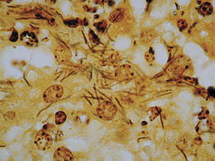
Acutely affected, recently weaned rabbitsmanifest profuse watery diarrhea, with mortality ranging from 15% to 50%. Survivors may be chronically affected, with depression, anorexia, weight loss, and cachexia. Necrotic and hemorrhagic enteritis are observed mainly in the ileum, cecum, and colon, with marked serosal andsubmucosal edema, congestion, and distinctreddening of cecal tissues. Multifocal hepatic necrosis generally appears after the acute phase of the disease in rabbits. Focal myocardial necrosismay also occur. Bundles of slightly gram-negative and silver-positive rod-shaped bacilli can be seen in viable hepatocytes bordering necrotic foci, in myocytes around necrotic foci in the heart, and in enterocytes and smooth muscle cells ofthe muscularis mucosa of the intestine. The occurrence of antibodies to C. piliforme in some species suggests that subclinical infection is common.


The disease in foals usually occurs before5 weeks of age, and in Arabian foals, combined immunodeficiency may play a role in susceptibility. Clinicopathologic abnormalities include leukopenia, hyperfibrinogenemia, metabolic acidosis, and hypoglycemia. Gross lesions include icterus, focal pale tan areas in the liver, and catarrhal enterocolitis. Focal, dark red lesions may be present in the small intestine, and the mesenteric lymph nodes may be enlarged and hyperemic. Multiple discrete and confluent foci of hepatic necrosis, hemorrhage, sinusoid congestion, infiltration of the portal triads with inflammatory cells, and bile duct hyperplasia are observed microscopically. Intestinal lesions may be mucohemorrhagic, and consist of mucosal necrosis with inflammatory cell infiltration and submucosal lymphoid hyperplasia. Hemorrhage, and necrosis of lymphoid follicles, is also present in spleen and mesenteric lymph nodes. As in the manifestation of the disease in other species, bacilli can be demonstrated in hepatocytes at the margin of liver lesions. The dams of affected foals may be carriers of the disease, and, judging by antiflagellar antibody titers, infection of horses may be quite common.


Rats with subclinical infections can transmit C. piliforme to naive rats. It seems likely that strains of C. piliforme display host specificity, and use of sentinels of one species may fail to demonstrate the presence of strains that are specific for another. Furthermore, certain strains of rats are more susceptible than others, which is a factor of major importance in colonies containing animals of multiple genotypes. Murine susceptibility varies with host strain, age, and immune status.


In dogs the infection is typically seen in the very young. Pups display widely disseminated lesions, with hepatitis, myocarditis, and enterocolitis. Disease in older dogs is less commonand may be related to an immunocompromised state.


Resistance to infection in mice is correlated with antibody titers, but neutrophils and natural killer cells may play an important role in the pathogenesis of murine Tyzzer’s disease. In juvenile mice, experimental depletion of either neutrophils or natural killer (NK) cells increases the severity of the disease, whereas in adults, NK cell depletion significantly increases the severity of disease in resistant (C57BL/6) but not in susceptible (DBA/2) mice. Macrophage depletion does not alter the course of infection.


Rats inoculated orally with spores of C. piliforme develop necrotic lesions in the intestines, liver, and heart during the first 2 weeks after inoculation, and infective spores are shed in feces. Rats that are clinically recovered can remain infected, with recrudescence of infections following steroid treatment.


Some isolates of C. piliforme are cross-infective (affecting more than one laboratory animal species) and others have a more limited host range. The reasons for this host specificity are not known. There are recent reports of cytotoxin production.

< div class='tao-gold-member'>

Stay updated, free articles. Join our Telegram channel

Jul 18, 2016 | Posted by in PHARMACOLOGY, TOXICOLOGY & THERAPEUTICS | Comments Off on The Genus Clostridium

Full access? Get Clinical Tree

Get Clinical Tree app for offline access