The Genus Bacillus

Chapter 7 The Genus Bacillus


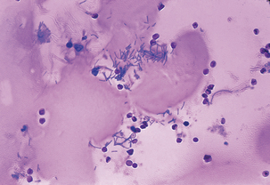
Bacilli are strictly aerobic or facultatively anaerobic, spore-forming rods (Figure 7-1). Most are catalase positive and motile, many are gram variable, and all are mediumto large. Spores may be readily visualized or may require induction by incubation at 42° C or for an extended period at 37° C.
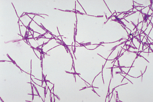


The Bacillus anthracis genome comprises an approximately 5.3 Mb chromosome and two plasmids, pX01 and pX02. The genome of at least two strains has been sequenced, and genetic analysis has focused mainly on the structural genes for toxin proteins (pagA, lef, and cya, located on pX01, see later) and the capsular biosynthetic genes (capB, capC, and capA, in a putative operon on pX02). The plasmids carry regulatory genes atxA (on pX01) and acpA (on pX02), which control toxin and capsule synthesis, respectively. A chromosomal gene, abrB, controls phase-specific transcription of the toxin genes.


Genetic exchange can occur among B. anthracis strains, as well as between B. anthracis andclosely related species; pX01 and pX02 are not self-transmissible, but can be transferred by conjugative plasmids of Bacillus thuringiensis. Bacillus anthracis is genetically indistinguishable from Bacillus cereus and B. thuringiensis; evolution of B. anthracis and B. thuringiensis from B. cereus, with subsequent gain of plasmid-based genes and loss of chromosomal genes or gene function may explain the emergence of the anthrax bacillus.


Anthrax in Domestic Animals


Anthrax continues to be reported among domestic and wild animals in the United States. The incidence of anthrax in U.S. animals is unknown;however, reports of animal infection have occurred among the Great Plains states from Texas to North Dakota and from eastern New York to California.


Signs of anthrax appear following a highly variable incubation period, which is usually 3 to7 days, but may be as short as 24 hours or greater than 2 weeks. The course of peracute anthrax in cattle and sheep may be as little as 1 to 2 hours, with sudden death a result of rapidly developing cerebral anoxia and pulmonary edema; fever (≤107° F), respiratory distress, and convulsions may be observed, but death is often reported as due to lightning strike. Epistaxis is common, and rigor mortis is often absent. One or two animals may die, followed by many others after several days. Acute anthrax in ruminants, with a clinical course of 24 to 48 hours, is characterized by an abrupt rise in temperature, anorexia, excitement followed by depression, and convulsions; in some cases there is evidence of pharyngeal involvement (lingual edema, with fluid accumulation in the throat and sternum) and respiratory distress. Animals may hemorrhage from the mouth, nose, and anus before death.


The course of equine anthrax is usually acute to subacute, with affected animals often surviving for 96 hours. Ingestion of spores results in septicemia, with enteritis and colic; disease following transmission by insect bite is initiated after the manner of human cutaneous anthrax, but the local subcutaneous edema spreads, often affecting the throat, ventral thorax, and abdomen. It is important to distinguish this syndrome from Corynebacterium pseudotuberculosis infection in the horse, which can also be associated with insect bites.


In the 1990s, B. anthracis was found during repair work in King’s Cross and Liverpool Street Stations in London. This should not have been surprising; the organism was associated with horse hair used in wall plaster in stations that were built in the nineteenth century. Decontamination is routine in these situations, and there is littleor no risk to the public. Similar situations were encountered in the past; use of saddle blankets woven from contaminated materials has led to equine anthrax, and manufacture of shaving brushes with B. anthracis–contaminated bristles resulted in cases of human cutaneous anthrax.


Omnivores and carnivores may have natural resistance to anthrax, and are more likely than other species to recover from the disease. Anthrax is typically subacute to chronic, and usually occurs after ingestion of contaminated meat or, in the case of swine, meat-and-bone meal. Lingual and pharyngeal edema, a common manifestation of infection in regional lymph nodes, may causedyspnea and dysphagia, with discharge of serosanguineous fluid from the mouth and death dueto asphyxia. Necrosis in the upper gastrointestinal tract, as well as mesenteric lymphadenopathy, occurs in carnivores. Cats may develop jowl and lingual carbuncles, cranial swelling, and severe gastroenteritis. An intestinal form of chronic anthrax has also been reported in swine.


Anthrax is not uncommon among wild carnivores. This is almost always associated with consumption of meat from animals dead of anthrax, sometimes when moribund, and presumably anthrax-affected, animals are shot as food forcarnivores in game farms. In these cases, anti-microbial therapy, as well as antimicrobialprophylaxis and immunoprophylaxis, carry the obvious risks associated with patient ingratitude.


Human Anthrax


Spores remain viable for decades, and, as such, soil is the usual source of infection for ruminants. Human infection following contact with contaminated soil usually takes the cutaneous form. Gastrointestinal and inhalation anthrax result from contact with products from affected animals, and, of course, may arise from laboratory exposure. Human anthrax is noncommunicable, regardless of the form of the disease.


Cutaneous anthrax (Figure 7-2) follows entry of the organism into a cut or abrasion. The incubation period is usually 24 to 72 hours, but can range up to 2 weeks. Signs begin with a painless papule, which becomes vesicular in 1 to 2 days, and can be surrounded by an extensive area of edema. The vesicle ulcerates by day 5 or 6 and the lesion dries, leaving a blackened necrotic area, the so-called black eschar. Affected individuals may also experience fever, malaise, headache, and swelling of draining lymph nodes. About 20% of untreated cases will progress to fatal septicemia. The case fatality rate with timely antimicrobial therapy is less than 1%, although treatment does not stop the progression of lesions.


Gastrointestinal anthrax follows consumptionof contaminated meat. A recent outbreak in Russia was associated with meat given to farm workers in lieu of wages; exposure of more than 1500 people led to 23 hospitalizations and 1 death. The incubation period of gastrointestinal anthrax is approximately 1 to 7 days. Pharyngeal lesions appear onthe base of the tongue and tonsils and are accompanied by sore throat, dysphagia, and regionallymphadenopathy. Inappetence, nausea and vomiting, and fever, followed by abdominal pain, vomiting of blood, and bloody diarrhea are signs ofacute colonic inflammation. Dissemination of B. anthracis from the initial site of infection leads to massive septicemia and toxemia. The case fatality rate is 25% to 60%, and even prompt antimicrobial therapy is not always sufficient to prevent mortality.


Cutaneous and gastrointestinal anthrax in humans may follow naturally occurring animal anthrax, but aerosol exposure is most likely to take place in laboratories, textile mills, or following a biological weapons attack. Pulmonary, or inhalation, anthrax results from respiratory exposure to spores. The infectious dose has been estimated variously as 8000 to 50,000 spores, and the incubation period is uncertain; recent experience suggests that it can be quite short (as little as 1-2 days), but some cases associated with the Sverdlovsk outbreakof 1979 (see Suggested Readings) were apparently as along as 43 days. The length of the incubation period may be inversely related to the dose. Spores do not all germinate immediately, but may in fact remain dormant until engulfed by pulmonary alveolar macrophages. This phenomenon is of particular importance in regard to duration of administration of therapy or prophylaxis; spores are not susceptible to antimicrobials, so these drugs must be present throughout the period in which germination may occur. Delayed onset does not appear to be an issue with cutaneous or gastrointestinal infections.


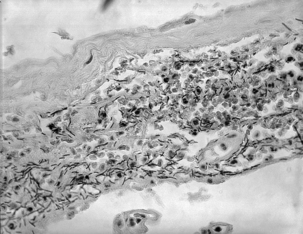
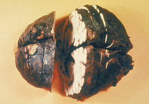
As spores germinate in macrophages and are carried to mediastinal lymph nodes, the patient may experience sore throat, mild fever, and muscle aches, which progress rapidly to severe respiratory difficulty. Organisms originating in the lungs and, later, those in the bloodstream, are temporarily removed by the reticuloendothelial system; however, B. anthracis ultimately escapes the lymphatics to produce overwhelming septicemia, usually with meningitis (Figures 7-3 through 7-5). Sudden onset of acute symptoms, including hypotension, edema, and fatal shock follow in2 to 5 days. Estimates of the case-fatality rate arebased upon limited data, but are probably at least90% and, without therapy, are more nearly 100%. Therapeutic intervention is of little use unlessinitiated quite early in the clinical course.


In the United States, as many as 130 cases of human anthrax occurred each year at the beginning of the twentieth century, but this declined to zero annual incidence through most of the 1990s. Most infections have been cutaneous, although 18 were pulmonary, the last in 1976 in a weaver. Worldwide, human anthrax is particularly common in agricultural regions with inadequate programs for control of livestock anthrax; in South and Central America, southern and eastern Europe, Asia, Africa, the Caribbean, and the Middle East, where animal anthrax occurs frequently, human cases are common and more than 95% are cutaneous. A recent epidemic in Zimbabwe comprised nearly 10,000 cases, with a case fatality rate of less than 2%.


Bacillus anthracis and Biological Warfare and Bioterrorism


Manufacture of biological weapons involving B. anthracis has been mentioned. It has, in fact, been a focus of biological warfare (BW) programs for decades, in the West before the establishmentof biological weapons treaties in the late 1960sand, by all indications, into the 1990s in the former Soviet Union.


Evidence of this is provided by the unusual anthrax epidemic that occurred in Sverdlovsk (Ekaterinburg), Russia, in April and May 1979. It was officially attributed to consumption ofcontaminated meat, but there was considerable international sentiment that it resulted from an accidental release of spores from a military microbiology facility in the city. Examination of prevailing winds during the period preceding the outbreak revealed that the pattern of deaths, humans and domestic animals alike, was correlated with what would have been the plume from the facility. Necropsies revealed lesions typical of inhalation anthrax, including hemorrhagic necrosis of thoracic lymph nodes and hemorrhagic medi-astinitis; gastrointestinal submucosal hemorrhagic lesions were nearly universal and mesenteric lymphadenitis occurred in a few cases. In most, edema was demonstrable adjacent to sites of infection and hemorrhagic meningitis indicated hematogenous dissemination. Subsequent investigation, which included testimony by defectors formerly involved in the illicit Soviet biological weapons program, established the reality of this epidemic, the occurrence of which is still officially deniedby the Russian government.


B. anthracis is a likely BW and biological terrorism (BT) agent because of the stability of its spores, transmission by the respiratory route, and the high mortality of the resulting disease. Release of 50 kg of spores upwind of a city of a half million people would cause an estimated 220,000 cases of anthrax, with 95,000 deaths.


Putative release of anthrax spores has, on several occasions before September 11, 2001, and on many occasions since, proven to be a hoax; law enforcement agencies dealt with an average of 1 or 2 cases per week through the summer of 2002. Targets have included abortion clinics, publicofficials, and others. Cornstarch and baby powder seem to be the most commonly chosen stand-ins for anthrax spores.

< div class='tao-gold-member'>

Stay updated, free articles. Join our Telegram channel

Jul 18, 2016 | Posted by in PHARMACOLOGY, TOXICOLOGY & THERAPEUTICS | Comments Off on The Genus Bacillus

Full access? Get Clinical Tree

Get Clinical Tree app for offline access