Chapter 39 The Genera Mycoplasma and Ureaplasma
Mycoplasmas and ureaplasmas, members of the family Mycoplasmataceae, arethe smallest prokaryotes and are related phylogenetically to members of the genera Clostridium, Streptococcus, and Lactobacillus. Members are distinct from other bacteria in their small genome size (0.58-1.35 Mb, which accounts for their limited metabolic options for replication and survival), and lack of cell wall synthesis (which places them in the class Mollicutes [soft skin] and renders them resistant to antimicrobials that interfere with cell wall synthesis). Mollicutes are of gram-positive lineage, but absence of a cell wall makes them unable to retain crystal violet/Gram’s iodine, so Gram staining is not part of the identification process.
THE GENUS MYCOPLASMA
Diseases and Epidemiology
Mycoplasma spp. infect a variety of animals, and some of diseases attributed to them have major economic impacts (Table 39-1). As a general rule, infections are spread among susceptible individuals through direct or droplet contact with oral, nasal, ocular, or genital secretions. Mycoplasmas are typically introduced into a group by the addition of clinically healthy carrier animals.
TABLE 39-1 Mycoplasma Species That Infect Domestic Animals
| Species | Host(s) |
|---|---|
| M. agalactiae | Goats, sheep: contagious agalactia |
| M. alkalescens | Cattle: arthritis, mastitis |
| M. bovigenitalium | Cattle: infertility, mastitis |
| M. bovis | Cattle: arthritis, mastitis, pneumonia, abortions, abscesses, otitis media, genital infections |
| M. bovoculi | Cattle: conjunctivitis |
| M. californicum | Cattle: mastitis |
| M. canadense | Cattle: abortions, mastitis |
| M. capricolum ssp. capricolum | Goats, sheep: mastitis, septicemia, polyarthritis, pneumonia |
| M. capricolum ssp. capripneumoniae | Goats: contagious caprine pleuropneumonia |
| M. conjunctivae | Sheep, goats: infectious keratoconjunctivitis |
| M. cynos | Dogs: pneumonia |
| M. dispar | Cattle: bronchiolitis |
| M. equigenitalium | Horses: abortion? |
| M. felis | Cats: conjunctivitis, pneumonia |
| Horses: pneumonia | |
| M. gallisepticum | Chickens, turkeys: airsacculitis, sinusitis |
| M. gatae | Cats: chronic arthritis, tenosynovitis |
| M. hyopneumoniae | Pigs: enzootic pneumonia |
| M. hyorhinis | Pigs: polyarthritis, polyserositis |
| M. hyosynoviae | Pigs: polyarthritis |
| M. iowae | Turkeys: embryo mortality |
| M. meleagridis | Turkeys: airsacculitis, skeletal abnormalities, decreased growth |
| M. mycoides ssp. capri | Goats: arthritis, mastitis, pleuropneumonia, septicemia |
| M. mycoides ssp. mycoides (small-colony type) | Cattle, domestic water buffalo: contagious bovine pleuropneumonia |
| M. mycoides ssp. mycoides (large-colony type) | Goats, sheep: mastitis, septicemia, polyarthritis, pneumonia |
| M. ovipneumoniae | Goats, sheep: pleuropneumonia |
| M. pulmonis | Laboratory rats and mice: murine respiratory mycoplasmosis |
| M. synoviae | Chickens, turkeys: infectious synovitis |
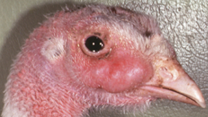
In poultry, the predominant mycoplas-mal pathogens are Mycoplasma gallisepticum, Mycoplasma synoviae, Mycoplasma meleagridis, and Mycoplasma iowae. Mycoplasma gallisepticum infection commonly results in chronic respiratory disease in chickens. Clinical signs include nasal discharge, coughing, sneezing, tracheal rales, and conjunctivitis. Turkeys are more susceptible than chickens and often develop severe sinusi-tis (Figure 39-1). Other less common diseasesyndromes are keratoconjunctivitis, arthritis, salpingitis, and encephalopathy. Mycoplasma synoviae usually causes a subclinical upper respiratory infection but may result in airsacculitis and synovitis in chickens and turkeys. Some strains of M. synoviae produce subclinical infection, a characteristic that creates difficulty in the management and control of disease outbreaks. Infectionsmay result in financial loss to the industry through processing condemnations, as well as reduced growth efficiency and egg production. Mycoplasma meleagridis causes respiratory disease in young turkeys and is involved in stunting, poor feathering, and leg problems. Mycoplasma iowae infection is economically important in turkeys, and the organism has occasionally been isolated from chickens. In turkeys it has been associated with reduced hatchability and embryo mortality, whereas experimentally infected chickens and turkeys develop airsacculitis and leg abnormalities. Avian mycoplasmas are egg transmitted and may spread laterally by direct or indirect contact. The mode of transmission of M. meleagridis to the egg is venereal. Infection of the male thallus results in semen contamination, which leads to oviduct infection after mating. With all of the avian pathogenic mycoplasmas, disease severity is dependent on the virulence of the strain involved, the age and breed of the bird, the degree of stress, methods of management, and presence of concurrent bacterial or viral infections.
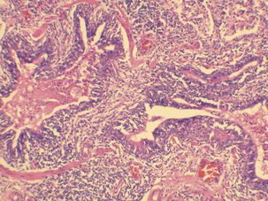
Three species of mycoplasmas commonly causing disease in swine are Mycoplasma hyopneumoniae, Mycoplasma hyosynoviae, and Mycoplasma hyorhinis. Mycoplasma hyopneumoniae is recognized as the etiologic agent of porcine enzootic pneumonia, which is characterized by high morbidity and low mortality that is commonly complicated by other opportunistic bacterial or viral infections. Enzootic pneumonia is an important economic problem affecting swine production worldwide. Clinical pneumonia is common in young animals but generally not seen in adults. Mycoplasma hyopneumoniae is transmitted from older to younger pigs by contact. Mechanical transmission by man and other animals is possible, and the organism may spread on the wind for several miles. Surveys conducted in different countries have revealed lesions of enzootic pneumonia in 30% to 80% of slaughter pigs. Feed conversion may be reduced by 14% to 20% and rate of gain by 16% to 30% in affected swine. The primary clinical sign is a sporadic dry nonproductive cough. Other signs may include fever, dyspnea, or impaired growth. Typical lesions are located in the apical and cardiac lobes of the lung, andconsist of well-demarcated, dark red to purple areas in acute disease, or tan to gray areas in chronic disease. Microscopic examination reveals characteristic lesions that include suppurative bronchopneumonia, histiocytic alveolitis with peribronchiolar and perivascular lymphohistiocytic cuffing, and nodule formation typical of bronchoalveolar lymphoid hyperplasia (Figure 39-2).
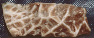
More than 20 species of Mycoplasma have been recovered from cattle. Many are purely commensal in nature, whereas others are responsible for a wide variety of clinical manifestations. Mycoplasma mycoides ssp. mycoides, isolated in 1898, was the first Mycoplasma species associated with animal disease. The small colony variant is the causative agent of contagious bovine pleuropneumonia (CBPP), one of the most important cattle diseases in history. The first veterinary college was founded to train practitioners to deal with CBPP. The disease has been eradicated from North America, Europe, and Australia, but remains endemic in parts of Africa and Asia. The disease is characterized by marked edema of the interlobular septa, with diffuse pneumonia and serofibrinous pleuritis (Figure 39-3). Infection spreads slowly within a herd, and may reach a peak morbidity rate of 50% only after several months. The disease may have acute or chronic manifestations.
Mycoplasma bovis is among the most virulent mycoplasmas of cattle worldwide. It is most often associated with bronchopneumonia, and infection is believed to be a predisposing factor inthe development of bovine respiratory disease complex, or shipping fever. Mycoplasma bovis is capable of systemic invasion, and arthritis and meningitis may be sequelae to mycoplasmemia. In addition, M. bovis has been associated with otitis media, mastitis, keratoconjunctivitis, endometritis, oophoritis, abortion, and seminal vesiculitis. In the United States alone, costs in the form of decreased weight gains and loss of production have been estimated at $32 million per year.
Pathogenesis
Cytadhesins are among the major virulence determinants of mycoplasmas. One such is the P97 protein of M. hyopneumoniae. The organism attaches to cilia of respiratory epithelial cells, resulting in damage to and subsequent loss of cilia (Figure 39-4). Lung damage results, as the lungs become predisposed to infection by secondary bacteria and the effects of other irritants. Cytadhesins have also been identified in M. gallisepticum, M. synoviae, and the human pathogen, M. pneumoniae.
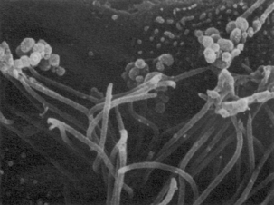
FIGURE 39-4 Mycoplasma hyopneumoniae associated with cilia in the porcine respiratory tract.
(Courtesy Eileen Thacker.)
Stay updated, free articles. Join our Telegram channel

Full access? Get Clinical Tree


