The Genera Chlamydia and Chlamydophila

Chapter 41 The Genera Chlamydia and Chlamydophila


Members of the order Chlamydiales are gram-negative obligate intracellular bacterial pathogens that parasitize hosts from humans to amebae. The family Chlamydiaceae has recently undergone extensive taxonomic revision, and the two proposed genera are Chlamydia and Chlamydophila. The genus Chlamydia would include Chlamydia trachomatis, Chlamydia suis, and Chlamydia muridarum. Six species assigned to the genus Chlamydophila are Chlamydophila abortus, Chlamydophila caviae, Chlamydophila felis, Chlamydophila pecorum, Chlamydophila pneumoniae, and Chlamydophila psittaci. This newly proposed nomenclature is not yet widely accepted.


Chlamydiae were once thought to be protozoa, and later were thought to be viruses because of their ability to pass through 450 nm filters. Ultimately, it has become clear that chlamydiae are bacteria. Molecular analysis of rDNA has confirmed them to be gram negative. Chlamydial cell walls contain cytoplasmic and outer membranes, but unlike other bacteria have no peptidoglycan layer. The cell wall also contains a genus-specific lipopolysaccharide. All chlamydiae encode an abundant major outer-membrane protein (MOMP), which is the principal determinant for serologic classification of isolates.


Some strains of C. psittaci and C. trachomatis are considered to be “energy parasites,” because host-derived ATP meets their energy needs. This may, in fact, be a requirement for all members of the family Chlamydiaceae.


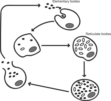
The order Chlamydiales has a developmental cycle (Figure 41-1) that is unique among prokaryotes, with two morphologically distinctive forms. The elementary body is the smaller (300-400 nm), infectious form, whereas the reticulate body is larger (800-1000 nm) and noninfectious. Elementary bodies are similar to spores, in that they are designed for survival in the environment. The reticulate body is metabolically active and capable of replication.


DISEASES AND EPIDEMIOLOGY


The Chlamydiaceae are pathogens of mammals, birds, and reptiles, and most have specific host or disease associations (Table 41-1). The host may serve as a natural reservoir of infection because many chlamydiae inhabit hosts in an asymptomatic state. Chlamydiae are spread by direct contact or through aerosols and do not require an alternate vector.


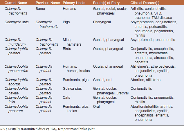
Chlamydia trachomatis is a human pathogen. Various strains are etiologic agents of prevalent sexually transmitted diseases and are the main causes of preventable blindness (trachoma) worldwide. Cervical infections, pelvic inflammatory disease, ectopic pregnancy, and infertility may affect women. Infants, infected in the birth canal, may develop pneumonia or conjunctivitis. In most cases, the conjunctivitis is self-limiting, but in some instances it results in blindness. In males, urethritis and lymphogranuloma venereum predominate. The latter condition is characterized by swollen lymph nodes in the groin. A secondary consequence of infection, more common in men than in women, is arthritis or Reiter’s syndrome. This organism has also been found in the temporomandibular joint (TMJ) of patients with TMJ disorder.


Diagnosis of swine chlamydial infections is relatively rare, especially in the United States, partly because most veterinary diagnostic laboratories do not routinely examine tissues for the presence of chlamydiae. The swine chlamydia C. suis were initially referred to as C. trachomatis, based on shared biologic characteristics; these organisms are susceptible to sulfadiazine and form glycogen-containing intracellular inclusions. Most infected animals remain asymptomatic. However, pneumonia, rhinitis, polyarthritis, conjunctivitis, pericarditis, and enteritis have been reported. Pre- and postweaning intestinal disease in pigs has been described, and affected animals have mild coughs, weight loss, and diarrhea. It is noteworthy that chlamydial infections have been detected in the intestines of clinically normal pigs and chlamydial antibodies have been identified in a high percentage of apparently healthy animals.


Chlamydia muridarum contains two strains, isolated from hamsters and mice, that previously belonged to C. trachomatis. The hamster strain may be avirulent, and murine infection may be inapparent or manifest as pneumonia.


Chlamydophila psittaci is primarily a pathogen of birds, with infections documented in more than 150 species worldwide. The disease has been given several names. Psittacosis is a disease of psittacine (parrot-type) birds. Ornithosis is the same disease affecting nonpsittacine birds. Other names used are avian chlamydiosis or parrot fever. Six serovars have been isolated from birds (A-F) and two from mammals (M56 and WC); virulence among serovars is variable. Serovar identification is useful for epidemiologic purposes. Most (50%-80%) of a flock will exhibit clinical signs when infection is caused by a highly virulent strain. Signs include anorexia, fever, conjunctivitis, production of yellowish green gelatinous droppings, and decreased egg production. Morbidity from less virulent strains is only 5% to 20%. Mortality attributed to highly virulent strains ranges from 10% to 30%, but is only 1% to 4% with less virulent strains. Lesions are similar, regardless of virulence, although they are less extensive with strains of lower virulence; airsacculitis, lung congestion, fibrinous pleuritis and/or pericarditis, hepatitis, splenomegaly, and enteritis are common. Recovered birds may shed chlamydiae in feces and nasal discharges for extended periods and pose a health threat to people.


Bacterial infection of humans with C. psittaci is also referred to as psittacosis. Infection usually occurs following inhalation of aerosolized bacteria from droppings, feathers, or tissues of infected birds. Transmission also may be mouth to beak, although cases acquired in this fashion are no doubt underreported, for obvious reasons. Most human disease results from exposure to infected cockatiels, parakeets, parrots, and macaws, and contact with birds through work or hobbies is a significant risk factor. The incubation period is 4 to 15 days, and the disease is a flulike illness with sudden onset of fever, chills, headache, weakness, and muscle aches. Some people also may have a dry cough, chest pain, breathlessness, and, in severe cases, pneumonia. Psittacosis in humans can be treated with antibiotics, and diagnosis is usually confirmed by blood tests and chest radiographs. Person-to-person transmission does not occur.


Three biovars of C. pneumoniae have been designated TWAR, Koala, and Equine. Biovar TWAR, derived from the laboratory designation of the first conjunctival and respiratory isolates (TW-183 and AR-39, respectively), is strictly a human pathogen. The organism causes bronchitis and pneumonia. Seroepidemiologic studies, direct detection of the organism within lesions, and isolation of the organism from atherosclerotic plaques have associated this strain with cardiovascular disease. Recently, researchers have suggested a connection to Alzheimer’s disease, which is based on the ability of C. pneumoniae to infect blood vessel endothelial cells and cause inflammation.


Biovar Koala strains have been isolated exclusively from ocular and urogenital sites of koalas, and do not appear to be highly virulent. The single strain of biovar Equine was isolated from the upper respiratory tract of a horse with a serous nasal discharge. Experimental inoculation of horses with this strain leads to asymptomatic infection.


Chlamydophila psittaci isolates associated with abortion in sheep, goats, cattle, and swine are now called C. abortus. These strains are endemic among ruminants and are colonizers of the placenta. Enzootic ovine abortion is the most common disease attributed to this organism, and is reported in the United States, South Africa, and Europe. The disease is characterized almost exclusively by a loss of lambs in late pregnancy, although weak or dead lambs may be born at term. Retained placenta and vaginal discharge are common clinical signs. Chlamydiae are present in high numbers in fetal membranes and in vaginal discharge; under these conditions, disease is spread to other ewes by the oral route. Bacteria can persist in ewe lambs for up to 2 years before causing abortion. The organism may have a broader than believed host range in that it has apparently been recovered from abortions in horses, rabbits, guinea pigs, and mice. Pregnant women working with infected sheep are at increased risk of abortion.


Chlamydophila caviae, which contains guinea pig strains formerly designated C. psittaci, is an agent of conjunctivitis. The infection is noninvasive, and attempts to infect other species of laboratory rodents have been relatively unsuccessful.


Feline strains of C. psittaci have been reclassified as C. felis. The organism is endemic among domestic cats worldwide and is a well-recognized cause of conjunctivitis, rhinitis, and, to a lesser degree, pneumonia. Of these syndromes, acute, chronic, or recurrent conjunctivitis is the most common clinical presentation. Experimental ocular inoculation of cats consistently results in acute conjunctivitis. Some experimentally infected animals develop persistent gastrointestinal or genital infections as a result of colonization of those mucosal surfaces. Whether chlamydiae are responsible for naturally occurring feline gastrointestinal or reproductive disease remains to be determined; however, infections at these sites may be a means by which disease is transmitted. Zoonotic infection has been linked to endocarditis, glomerulonephritis, chronic cough, and flulike illness in humans.


Chlamydiae isolated from mammals, but not associated with a specific host, have been assigned to the species C. pecorum. This organism causes infertility and genitourinary disease in koalas. Sporadic bovine encephalomyelitis, also known as Buss disease, affects young cattle and buffaloes, and is characterized by encephalitis, peritonitis, and fibrinous pleuritis and occurs worldwide. The incubation period is 1 to 4 weeks, and affected animals are anorectic and febrile. Hypersalivation, dyspnea, and ataxia are frequently observed, and mortality is 50%. Organisms are located in the brain, spinal cord, and lymph nodes, indicating systemic infection. Enteritis and arthritis in swine and sheep have occasionally been attributed to infection with C. pecorum.


Chlamydiosis has been described in chameleons, turtles, tortoises, crocodiles, and snakes. Manifestations include pericarditis, hepatitis, enteritis, pneumonia, and splenitis. Most of these strains remain uncharacterized.

< div class='tao-gold-member'>

Stay updated, free articles. Join our Telegram channel

Jul 18, 2016 | Posted by in PHARMACOLOGY, TOXICOLOGY & THERAPEUTICS | Comments Off on The Genera Chlamydia and Chlamydophila

Full access? Get Clinical Tree

Get Clinical Tree app for offline access