The Family Anaplasmataceae

Chapter 40 The Family Anaplasmataceae


The family Anaplasmataceae has beenreorganized based on 16S rRNA gene sequences to include all species ofα-Proteobacteria in the genera Neorickettsia, Anaplasma, Ehrlichia, and Wolbachia; Aegyptianella has been retained as a genus incertae sedis (Table 40-1). Wolbachia spp. are associated exclusively with invertebrates, and the genera Eperythrozoon and Haemobartonella have been transferred to the order Mycoplasmatales. The obligately intracellular Anaplasmataceae replicate within cytoplasmic vacuoles of host cells, such as erythrocytes, reticuloendothelial cells, bone marrow–derived phagocytic cells, endothelial cells, and reproductive tissues of insects, arthropods, or helminths. Most members have a trematode, tick, or other invertebrate vector host.


THE GENUS NEORICKETTSIA


Neorickettsia spp. are small, nonmotile, coccoidto crescentric, intracytoplasmic, gram-negative bacteria that are found within vacuoles of monocytes, macrophages in lymphoid tissues, and occasionally in enterocytes. They stain readily with Giemsa or Macchiavello stains and cannot be cultivated in cell-free media or in chicken embryos. Flukes serve as vectors, and all stages within the life cycle of flukes are infectious. The infectious cycle includes transovarial and transstadialtransmission.


Neorickettsiae maintain their individual vacuolar membranes when they undergo binary fission, so intracellular inclusions may not be observed microscopically. Cell lysis leads to release of cell-free bacteria that can infect other host cells.


Species of note are N. helminthoeca, N. (Ehrlichia) risticii, and N. (Ehrlichia) sennetsu. Molecular and antigenic analyses have recently established a close relationship among these organisms and further similarities may exist in their modes of transmission.


Neorickettsia helminthoeca


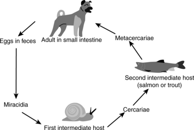
Salmon poisoning disease was first recognized in the early nineteenth century by settlers in the Pacific Northwest when their dogs fell ill after consuming raw salmon. The causative agent, N. helminthoeca, was characterized in 1953, but could not be placed in a previously described genus based on the disease and morphologic characteristics. It is unique, in that it is the only obligately helminth-borne pathogenic bacterium. Canine mononuclear cells are infected after dogs ingest salmonid fish encysted with a fluke, Nanophyetus salmincola (Figure 40-1), infected with the organism. The disease is indigenous to river areas of the U.S. Pacific Northwest coast, from northern California to southwestern Washington State. Occasional cases associated with fish migration have been reported in British Columbia.


The raccoon and spotted skunk are the principal definitive hosts for the fluke. Adult flukes develop in the intestinal lumen within 6 days after ingestion in dogs, cats, foxes, bears, coyotes, opossums, otter, mink, lynx, and birds. Infected eggs shed in feces hatch and release a miracidium. The miracidium penetrates the first intermediatehost, the snail Oxytrema silicula, where the cecariae (free-living trematode larvae) develop. Cercariae are liberated and penetrate the skin of the secondary intermediate host, which are fish, usually of the family Salmonidae, or Pacific giant salamanders. Metacercariae are found in the muscle, kidney, eye, and other organs of fish and salamanders. Neorickettsia helminthoeca is maintained by transovarial passage in the helminth and is found throughout the life cycle of the fluke, including free-swimming cercariae.


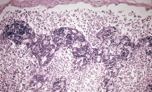
Salmon poisoning (Figure 40-2) generally affects only members of the family Canidae, but death of captive polar bears has been reported in association with salmon poisoning disease. After ingestion of infested fish, the trematodes attach to and penetrate deeply into the mucosa, particularly in the duodenum but also throughout the small and large intestines. The precise mechanism of infection remains to be elucidated, but superficial enteritis develops rapidly, and progresses to hemorrhagic enteritis. Bacteria spread by the hematogenous route to lymph nodes, spleen, liver, lungs, brain, and thymus. Acute disease is characterized by fever, depression, dehydration, anorexia, diarrhea, and lymphadenopathy. The case fatality rate in untreated dogs is 50% to 90% and the exact cause of death remains unknown. Recovered animals are immune to reinfection.


Difficulties related to bacteriologic culture of N. helminthoeca have been a detriment to studying disease pathogenesis. Recently propagation in a continuous cell line has facilitated antigenic and genetic analyses. Canine monocytes, canine leukocytes, mouse lymphoblasts, and a macrophage line support in vitro growth.


Antemortem diagnosis is based on detection of fluke eggs in feces, presence of compatible clinical signs, history of travel to the Pacific Northwest, demonstration of rickettsiae in lymph node aspirates, and serology. Trematode eggs are operculated and are passed in the feces 5 to 8 days after fish ingestion. They can be detected by direct smears or sugar flotation techniques. Macchiavello or Giemsa stains of lymph node aspirates reveal intracytoplasmic rickettsial bodies, but organisms are not detected by microscopic examination of circulating lymphocytes. Affected animals typically seroconvert 13 to 15 days postinfection, so several veterinary diagnostic laboratories offer serologic diagnosis by way of indirect fluorescent antibody or complement fixation tests.


Postmortem diagnosis is based on compatible gross and microscopic lesions. Changes areprimarily associated with lymphoid tissues, and include lymphadenopathy, splenomegaly, and petechiation of the gallbladder, pancreas, and mucosae of the esophagus, urinary bladder, and intestinal tract. General lymph node enlargement is attributed to marked infiltration of macrophages. Microscopic lesions include depletion of mature lymphocytes from lymph node cortex and medulla, nonsuppurative meningoencephalitis, splenic follicular central hemorrhage and necrosis, and obliteration of thymic architecture. Coccoid bodies may be found within the macrophages.


Prevention of the disease involves not allowing raw or improperly cooked fish to be fed to dogs. Thorough cooking, or freezing at −20° C for24 hours, kills N. helminthoeca and metacercariae. If infected raw fish is eaten, apomorphine should be administered as an emetic. Sick animals are given supportive care to control vomiting and diarrhea, and to maintain acid-base balance. Parenteral administration of tetracyclines is usually helpful, but there are no commercial vaccines.


Neorickettsia (Ehrlichia) risticii


Neorickettsia risticii is the agent of Potomac horse fever (PHF), an acute diarrheic illness of equids, also known as equine monocytic ehrlichiosis or equine scours, because of the affinity of the organism for blood monocytes, tissue macrophages, and intestinal epithelial cells. The disease was first recognized in 1979 in areas bordering the Potomac River in Virginia and Maryland. Since then it has been diagnosed in most states within the continental United States, several Canadian provinces, and South America (Brazil, Venezuela, and Uruguay). Serologic evidence of PHF has been found in France, India, and Australia.


The epidemiology of PHF has remained obscure since its discovery more than 20 years ago. The disease appears to be restricted to low-lying regions in proximity to bodies of water, and there is a seasonal pattern with peak incidence occurring during summer. Arthropod vectors do not appear to be involved in transmission, but N. risticii is found in the feces of infected horses. Oral transmission has been demonstrated experimentally, but this route is not natural.


Neorickettsia risticii appears to be maintained in nature in a complex aquatic ecosystem. The infection cycle apparently involves an intermediate snail reservoir and a trematode cercarial vector. In the laboratory, researchers have infected mice and horses by intraperitoneal subcutaneous inoculation, respectively, with trematodes from snails. DNA of N. risticii has been found in virgulatecercariae from the freshwater snails Juga yrekaensis in northern California and Elimia livescens in Ohio. Such DNA has also been detected in metacercariae from aquatic insects, such as caddisflies, stoneflies, damselflies, mayflies, and dragonflies. Oral transmission of PHF has been demonstrated in horses fed caddis flies, and it is natural to speculate that transmission to horses involves accidental ingestion of insects harboring infected metacercariae. Potential helminth vectors include Lecithodendrium and Acanthatrium spp. These N. risticii–infected helminths have been found in the intestinal tracts of bats and birds, but no definitive reservoir host has been identified. Besides horses, other susceptible mam-mals include cattle, mice, dogs, and cats. In endemic locations, antibody titers to N. risticii have been found in goats, pigs, cats, dogs, and coyotes.


The primary clinical sign is acute, waterydiarrhea. Mild colic, anorexia, fever, depression, edema, dehydration, laminitis, and leukopenia are additional findings. Rarely, abortion may result from infection in the unborn fetus. The case fatality rate is 5% to 30%. Fatalities result if severely affected horses are not treated promptly with electrolytes, fluids, and appropriate antimicrobial agents.


The watery diarrhea is a direct result of enterocyte infection. Degeneration of infected epithelial cells, with loss of microvilli, results in accumulation of cAMP and physical inability to conduct electrolyte transport in the colonic mucosa. Malabsorption of sodium and chloride ions and lack of water resorption lead to diarrhea.


A diagnosis of PHF is suggested in horses exhibiting nonspecific signs and diarrhea during the summer months in areas endemic for N. risticii. Several options are available for the diagnosis of PHF, including an indirect fluorescent antibody test. Rising titers with accompanying clinical signs suggests active infection. Detection of the organism in infected tissues has been accomplished using a modified Steiner silver stain, an immunoperoxidase method, or transmission electron microscopy. Definitive diagnosis of PHF formerly required isolation of N. risticii in cell culture, but polymerase chain reaction (PCR) amplification of DNA from peripheral blood or feces has proven to be a highly specific and sensitive means of diagnosis.


Commercial, inactivated vaccines for PHF are available but the antibody response to vaccination has been poor and there are consistent reports of vaccine failure in the field, particularly in endemic areas. Heterogeneous strains of N. risticii have been recovered from field-vaccinated horses suffering from clinical PHF. Thus it appears that both a deficiency in antibody response and heterogeneity among isolates are responsible for the present vaccine failures. Infections can be treated by intravenous administration of oxytetracycline early in the course of the disease.


THE GENUS ANAPLASMA


With the inclusion of the former Ehrlichia bovis, Ehrlichia phagocytophila, Ehrlichia equi, and Ehrlichia platys, the description of the genus Anaplasma was amended to include gram-negative, small coccoid to ellipsoid bacteria that live within cytoplasmic vacuoles of myeloid cells, neutrophils, and erythrocytes in peripheral blood, or in tissues of the mononuclear phagocyte organs. Organisms occur singly, or more commonly in inclusions called morulae, and stain bluish purple by Romanovsky methods. Biologic vectors are usually ticks. The genus now consists of species Anaplasma bovis, Anaplasma caudatum, Anaplasma centrale, Anaplasma marginale, Anaplasma ovis, Anaplasma phagocytophilum, and Anaplasma platys.


Anaplasma marginale


Initially described by Theiler in 1908 as a developmental stage of the protozoan Babesia bigemina, A. marginale is the primary cause of bovine anaplasmosis, an economically significant disease on six continents. Biologic transmission occurs when infected hard ticks in the genera Boophilus, Dermacentor, Ixodes, or Rhipicephalus feed on immunologically naive cattle. Mechanical transmission occurs through biting flies or blood-contaminated fomites such as used hypodermic needles or dehorning instruments. Cattle that recover from acute infection remain persistently infected for years, with microscopically undetectable levels of the organism. Carriers develop clinical disease when stressed. Persistence in primary hosts is fundamental to continued transmission because transovarial transmission of A. marginale in the tick vector does not occur. Anaplasmosis is enzootic in the southern Atlantic and Gulf Coast states, on the lower plains, andin western states, but occurs sporadically in northern states. Natural and biologic vectors are seasonal, so there is a correlation between disease outbreaks and vector seasons.


Anaplasma marginale causes fever, anorexia, weight loss, decreased fertility in bulls, and icterus. Acute disease is characterized by severe anemia, pale mucous membranes, and lethargy. Animals may be belligerent if hypoxia affects the brain. Cows in late pregnancy may abort. Animals less than 1 year old are susceptible to infection but relatively resistant to disease, whereas cattle older than 36 months of age usually experience severe disease with a 30% to 50% case fatality rate.


Anaplasmosis induced by A. marginale occurs in four stages. The incubation period comprises the time from introduction of the agent into a susceptible animal until 1% of the red blood cells are parasitized. This varies from 3 to 8 weeks and depends on the initial number of infecting organisms. During this time the animal remains asymptomatic. The first clinical signs become apparent during the developmental stage, when more than 15% of the erythrocytes are infected. Fever is the initial finding, followed by anorexia, depression, and lethargy. The length of this period ranges from 4 to 9 days. The convalescent stage varies greatly in length, from weeks to months, and extends from the appearance of reticulocytesin peripheral circulation until the blood values return to normal. Mortality may occur duringthe early convalescent stage. Necropsy findings are attributable to hemolytic anemia. Grossly, the blood appears thin and watery. Other findings include tissue pallor, icterus, hepatosplenomegaly, and enlarged gallbladder. The carrier stage may last for the remainder of the life of the animal.


Within the tick, the bacterium undergoes a complex developmental cycle that involves the gut initially and ultimately the salivary gland, from which transmission occurs during feeding. When the organism invades mature erythrocytes, replication occurs by binary fission within membrane-bound cytoplasmic inclusions and two to eight infective initial bodies are formed. These leave the red blood cells by exocytosis to infect other susceptible erythrocytes.


The severity of the disease is related directly to the proportion of the erythrocyte mass destroyed. Hemoglobinuria does not occur in anaplasmosis because the destruction of erythrocytes occurs intracellularly rather than intravascularly. Serum factors sensitize erythrocytes to phagocytosis by the monocyte-macrophage system and these opsonins increase in the circulation before the hemolytic crisis. Fetal hypoxia is responsible for abortion.


Persistence of A. marginale in fully immunocompetent hosts is mediated by antigenic variation in the organism’s major surface proteins. Recently, A. marginale has been propagated in continuous culture in a tick embryo cell line. This system has been used as an infection model and for adhesion studies. Adhesion, infection, and transmission are mediated by major surface protein 1a.


A presumptive diagnosis of anaplasmosis is based on clinical signs and hematologic findings in animals in an endemic area. Serologic testing, direct examination of blood smears, and molecular methods are also useful. Rapid card agglutination, complement fixation, and enzyme immunoassays are available for serologic diagnosis. Phenotypic criteria for identification of ruminant erythrocytic Anaplasma spp. (A. centrale, A. marginale, and A. ovis) have for many years relied on subjective methods, such as the location of inclusion bodies in host red blood cells (Figure 40-3) and host pathogenicity (cattle vs. sheep). Recentstudies have confirmed the suitability of 16S rDNA sequence analysis to define the species from blood samples.
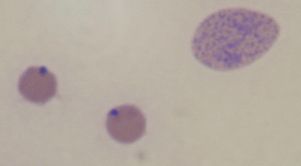

< div class='tao-gold-member'>

Stay updated, free articles. Join our Telegram channel

Jul 18, 2016 | Posted by in PHARMACOLOGY, TOXICOLOGY & THERAPEUTICS | Comments Off on The Family Anaplasmataceae

Full access? Get Clinical Tree

Get Clinical Tree app for offline access