CHAPTER 9 The Eyes and Associated Structures
GENERAL CONSIDERATIONS
Microscopic Evaluation
When identifying cell types, it is important to look in an area where individual cells can be evaluated. However, thick collections of material, often consisting of clustered epithelial cells or necrotic material, tend to be understained, and cells with granules that stain more readily than other components (mast cells and eosinophils), naturally pigmented elements (melanin), bacteria, and fungal hyphae may be visualized within or on top of the thick tissue (Figure 9-1). Inclusions found in epithelial or inflammatory cells may be normal elements, artifacts of treatment, or evidence of the pathologic process or etiology (Table 9-1). Normal tissue also may be present.
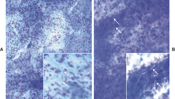
Table 9-1 Inclusions In or On Cells from Ocular Tissue
| Inclusions | Significance |
|---|---|
| Inclusions in or on epithelial cells | |
| Melanin granules | Normal in pigmented tissue Small granules may be confused with Mycoplasma organisms. |
| Mucin or mucin granules | Normal goblet cells |
| Surface mixed bacteria | Contaminants |
| Drug inclusions | Artifact of treatment with topical ophthalmic ointments |
| Mycoplasma spp. | Pathogen |
| Chlamydophila spp. | Pathogen |
| Neutrophils | Intact neutrophils within squamous cells: no known significance |
| Inclusions in neutrophils | |
| Bacteria | Pathogen |
| Small fungal organisms (e.g., Histoplasma spp.) | Pathogen |
| Pyknotic nuclei | Aging change or accelerated apoptosis |
| Inclusions in macrophages | |
| RBCs (erythrophagia) | Hemorrhage |
| WBCs (leukophagia): whole or degraded | Long-standing inflammation |
| Iron pigment (macrophages are termed hemosiderophages) | Chronic or previous hemorrhage |
| Melanin (macrophages are termed melanophages) | Pigmented tissue with release of melanin from ruptured or degraded epithelial cells |
| Certain bacteria (e.g. Mycobacterium spp.) | Pathogen |
| Some fungal organisms (e.g. Histoplasma spp.) | Pathogen |
| Protozoal organisms (e.g. Leishmania spp.) | Pathogen |
Once a category is identified, a more specific diagnosis may be possible. For example, a search for an etiologic agent is indicated if inflammation is present. At the very least, the category can guide additional testing or therapy. Special cytologic features of neutrophils, epithelial cells, and extracellular material are listed in Table 9-2 and often provide additional information about the pathologic process; misinterpretation of these features (e.g., mistaking free mast cell granules for bacterial cocci) can lead to erroneous conclusions.
Table 9-2 Special Cytologic Features and Their Significance
| Cytologic Feature | Significance |
|---|---|
| Neutrophils | |
| Nondegenerate: well-lobulated condensed nuclei, intact nuclear and plasma membranes | Neutrophilic or purulent inflammation: septic or nonseptic |
| Degenerate: swollen hypolobulated nuclei, fragmented nuclear or cytoplasmic membrane | Septic inflammation likely |
| Pyknotic: shrunken, condensed, rounded, and disconnected nuclear lobes | Aging change or accelerated apoptosis |
| Intracytoplasmic bacteria | Usually pathogen(s) |
| Epithelial Cells | |
| Dysplastic change: nuclear:cytoplasmic asynchrony | Secondary to inflammation; differentiate from epithelial neoplasia with secondary inflammation |
| Cornification/keratinization: keratin does not stain with Romanowsky stains; its presence is inferred when squamous cells are angular or folded | Abnormal for corneal epithelial cells; occurs in keratitis |
| Extracellular material | |
| Bacteria | Possible contaminants, but may be significant especially if found in corneal samples or if many bacteria of a single morphology are noted |
| Fungal organisms: yeast forms of Blastomyces, Cryptococcus, Coccidioidomyces, Histoplasma; hyphae of Aspergillus and other fungi | Pathogens |
| Parasites: larvae rarely seen cytologically | Pathogens |
| Free eosinophil, mast cell, or melanin granules | Indicate presence of ruptured eosinophils, mast cells, or epithelial cells; granules may be mistaken for bacteria |
| Cell fragments, especially stringy nuclear chromatin | Artifact of slide preparation; can resemble hyphae when surrounded by mucus |
| Cholesterol crystals | Epithelial degeneration |
| Stain | Artifact; may be mistaken for bacterial cocci |
| Mucus | Normal in areas where goblet cells are located; may be increased with some pathologic processes |
THE EYELIDS
Blepharitis
Blepharitis may be focal or diffuse and acute or chronic. Causes may be bacterial, mycotic, parasitic, allergic, or immune mediated. The objectives in cytologic examination of blepharitis are to characterize the type of exudate (neutrophilic, lymphocytic-plasmacytic, eosinophilic, or granulomatous) and search for the causative agent. Agents that may be encountered in scrapings are Sarcoptes spp. Demodex spp. dermatophytic yeast, and bacteria. Demodex folliculorum causes minimal exudation. Bacterial blepharitis, particularly staphylococcal, has a neutrophilic exudate. Certain fungi, such as Blastomyces dermatitidis, cause either a primarily neutrophilic or a pyogranulomatous exudate, whereas others cause a granulomatous exudate (macrophages, including epithelioid forms, and giant cells). Foreign bodies can elicit a pyogranulomatous or granulomatous response (Figure 9-2).
Immune-mediated disease usually is characterized by a neutrophilic exudate, but eosinophilic types also occur. The presence of either bacteria or a primarily neutrophilic exudate does not exclude allergic and immune-mediated causes, especially if the lesion is ulcerated. In cats, eosinophilic plaques may be manifested as periocular blepharitis.1 A fine-needle aspirate reveals primarily eosinophils, some mast cells, and a mixture of other white blood cell (WBC) types.
Discrete Masses
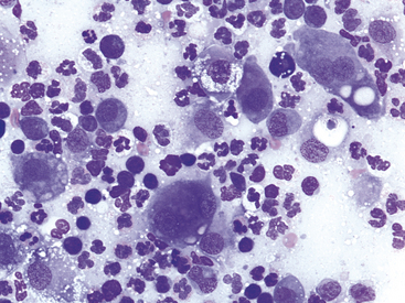
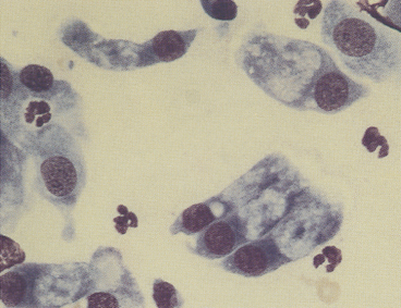
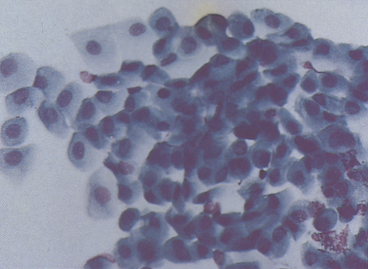
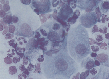
Discrete masses on the eyelids may be neoplastic (benign or malignant) or nonneoplastic. Among neoplasms, benign sebaceous gland tumors (sebaceous adenoma, sebaceous epithelioma) are the most common type on canine eyelids.2 The glands of Zeis and Moll at the eyelid margin and the meibomian glands, which lie beneath the palpebral conjunctiva and open at the lid margin, are all of the sebaceous type; tumors arising from them are similar to cutaneous sebaceous gland tumors. The cells are readily recognized by their voluminous vacuolated cytoplasm that nearly obscures small rounded nuclei (Figure 9-3). The malignant counterpart of these tumors is rare on the eyelids.
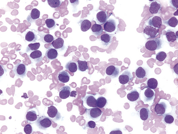
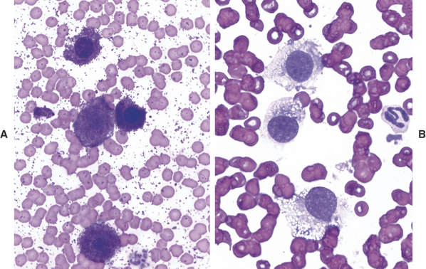
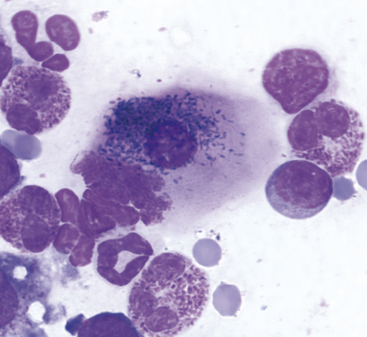
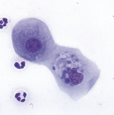
Other tumors frequently encountered on the eyelids and readily diagnosed by cytologic examination include melanoma (benign and malignant), histiocytoma (Figure 9-4), lymphoma, mast cell tumor (low and high grade) (Figure 9-5), papilloma, and squamous cell carcinoma. Squamous cell carcinomas are frequently ulcerated. In mast cell tumors, mast cell granules sometimes are not visible if quick stains are used (Figure 9-5, B). Other carcinomas and connective tissue tumors (fibrosarcoma, hemangiosarcoma, histiocytic sarcoma) occur less frequently and are discussed in Chapter 2.2,3
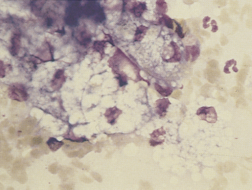
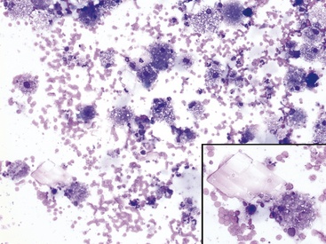
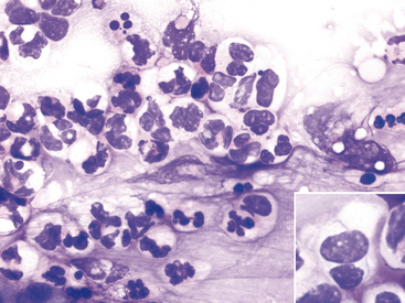
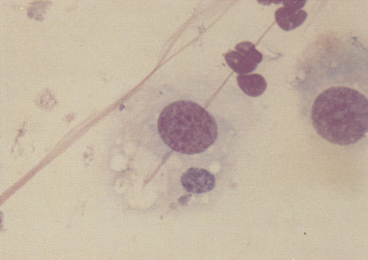
Nonneoplastic discrete masses unique to the eyelid include the hordeolum, a localized purulent lesion of sebaceous glands, and the chalazion, a lipogranuloma of the meibomian gland. Fine-needle aspiration of these lesions yields numerous foamy macrophages and a few giant cells and lymphocytes. The macrophages are apparently phagocytosing glandular secretory product; cytophagia is not prominent. Variable numbers of sebaceous epithelial cells also are found (Figure 9-6). Differentiating a hordeolum or chalazion from sebaceous gland adenoma by cytologic examination may be difficult if the latter has internally ruptured and caused secondary inflammation. Hordeolum or chalazion may contain inspissated secretory product or mineralized debris that appears as amorphous granular material on cytologic preparations.
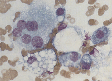
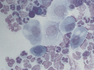
Idiopathic ocular adnexal granulomas may simulate neoplasms, be bilateral, and be a component of systemic granulomatous disease.4 Systemic histiocytosis of Bernese mountain dogs causes periocular granulomatous masses.5,6 True cysts can occur on the eyelids and typically contain foamy macrophages and cholesterol crystals from epithelial degeneration (Figure 9-7).
THE CONJUNCTIVA
The primary indications for conjunctival cytologic evaluation are to characterize an exudate and to attempt to identify the cause of conjunctivitis.7 Certain anatomic structures affect the types of cells found on all preparations from normal and diseased eyes. The conjunctiva is composed of two continuous layers of epithelium that lie in apposition. The inner epithelial layer of the eyelid, called the palpebral conjunctiva, is composed of pseudostratified columnar epithelium and interspersed goblet cells (Figure 9-8). Cilia may be found on the columnar cells. At the fornix, deep within the conjunctival sac, the epithelium reflects back over the globe. This bulbar conjunctiva is composed of stratified squamous epithelium (Figure 9-9). Bulbar conjunctiva is continuous with the corneal epithelium at the limbus. The squamous cells are noncornified and often contain melanin granules (Figure 9-10). In most conjunctival scrapings, squamous cells are more numerous than columnar cells.
In animals treated with topical ophthalmic ointments (particularly neomycin), epithelial cells may contain dense basophilic homogeneous cytoplasmic inclusions (Figure 9-11).8 Such inclusions must be differentiated from infectious agents. At the fornix, conjunctival lamina propria contains lymphoid tissue; various types of lymphoid cells may be found in any conjunctival scraping. Without clinical signs of conjunctivitis, little emphasis should be placed on the observation of lymphocytes or plasma cells among epithelial cells.
Cytologic preparations from the conjunctiva should include freshly derived cells. If external debris within the conjunctival sac is present, imprints of the debris should be made because this material may contain the etiologic agent, such because Blastomyces spp. More often, the debris obscures the primary lesion; therefore, after imprints are made the debris should be removed and conjunctival scraping performed with a flat, round-tipped spatula. Preparation of bulbar conjunctival imprints using filter strips following topical anesthesia has been reported in dogs.9
Neutrophilic Conjunctivitis
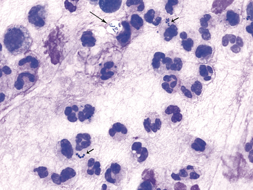
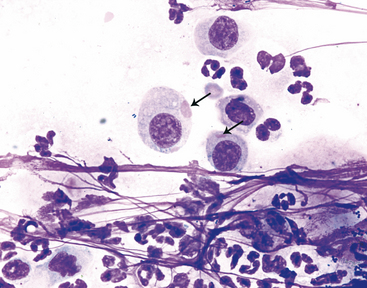
Canine and feline conjunctivitis frequently is neutrophilic and occurs with bacterial, viral, allergic, or other causes. Pseudomembranous (ligneous) conjunctivitis is neutrophilic.10 Cytologic evaluation may not reveal the cause. Neutrophils may be nondegenerate or degenerate (Figure 9-12). In cats, the latter are rarely encountered. In both species, intact neutrophils may be found within squamous cells (Figure 9-13), and the significance of this finding is unknown. Mucus is a common component of neutrophilic exudates and may cause cells to appear in rows on the smear.
The exudate of canine neutrophilic conjunctivitis often contains bacteria, regardless of primary cause. The bacteria are often large and/or small cocci and less frequently rods (Figure 9-14). The dilemma is whether the bacteria are of primary importance or are merely opportunistic. Normal bacterial flora of the canine conjunctival sac have been described.11 Keratoconjunctivitis sicca is a common canine disorder causing neutrophilic exudate in which bacteria frequently are encountered. The disease is diagnosed readily by the Schirmer tear test. In contrast to that of dogs, the exudate of feline neutrophilic conjunctivitis rarely contains bacteria. When observed, bacteria should be considered clinically significant in feline conjunctivitis.
Distemper is the most important viral cause of canine neutrophilic conjunctivitis. Canine distemper is diagnosed by its classic clinical signs and fluorescent antibody staining of conjunctival smears. Canine distemper inclusion bodies in epithelial cells are found rarely (Figure 9-15), and a search for them has limited diagnostic value.
A common cause of feline neutrophilic conjunctivitis is herpesvirus infection (Figure 9-16). Diagnosis is confirmed by a PCR assay,12,13 fluorescent antibody staining of conjunctival smears, or viral isolation. Multinucleated epithelial cells may be found, but intranuclear inclusion bodies rarely are seen.7,14
Neutrophils predominate also in the conjunctival exudate of feline chlamydial infection. In experimental Chlamydophila felis infections, organisms were found by postinoculation on day 6 when clinical signs first appeared.15 Solitary, large (3 to 5 μm), basophilic particulate forms initially are found in the cytoplasm of squamous epithelial cells (Figure 9-17). The particulate nature of the initial body is an important observation to distinguish C. felis from incidental foci of homogeneous cytoplasmic basophilia found in squamous epithelial cells (see Figure 9-11).8 Organisms also may appear as aggregates of coccoid basophilic bodies (elementary bodies), 0.5 to 1 μm in diameter (Figure 9-18). In the experimental infections, organisms rarely were found by day 14 postinoculation,15 and intracytoplasmic organisms are present only infrequently in chronic conjunctivitis.7 Chlamydial conjunctivitis may be confirmed by PCR or fluorescent antibody staining. Chlamydiae other than C. felis also may play a role in ocular disease in cats.16
Stay updated, free articles. Join our Telegram channel

Full access? Get Clinical Tree


