Chapter 10
The External Ear Canal
Anatomy of the External Ear
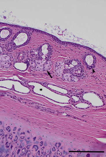
The external ear consists of cartilage and the overlying skin, which create the pinna and external acoustic meatus (between the base of the pinna to the tympanic membrane). The auricular cartilage determines the shape and appearance of the pinnae and supports the vertical ear canal. The annular cartilage, found at the base of the auricular cartilage, supports the horizontal and external ear canal. The skin covering the cartilage within the canal contains sebaceous glands, tubular ceruminous glands, and small hair follicles (Figure 10-1).1
Etiology and Pathogenesis of Otitis Externa
Otitis externa, inflammation of the skin and adnexal structures of the ear canal, is commonly encountered in veterinary patients. It is estimated to affect 10% to 20% of canines and 2% to 6% of felines presented for veterinary care.2,3
Primary Factors
Primary factors are those which initiate the inflammation of the external ear canal and include parasites, allergic skin disease, foreign bodies, disorders of keratinization, autoimmune diseases, trauma, sebaceous adenitis, zinc-responsive dermatoses, juvenile cellulitis, and certain endocrine disorders (Box 10-1).4–6
Predisposing Factors
Predisposing factors facilitate the development of otitis externa by promoting an environment suitable for the survival of the perpetuating factors. Predisposing factors not only include factors such as ear conformation, hypertrichosis of the ear canal; and breed predispositions, which are congenital, environmental, or both, but also iatrogenic trauma, excessive moisture, and obstructive ear disease (Box 10-2).3,4
Perpetuating Factors
Rather than initiating the otitis externa, perpetuating factors sustain the established disease; once the ear canal has been altered by primary and predisposing factors, opportunistic infections and progressive changes occur to prevent resolution of disease. These factors include bacteria, yeast, otitis media, and progressive hyperplastic changes of the ear canal caused by disease (Box 10-3).3,4
Cytologic Evaluation of Ear Canal Secretions
Collection and Staining of Samples
Samples of the ear canal secretions for cytologic evaluation are collected using separate cotton-tipped swabs for each ear canal. Samples should be collected after performing otoscopic examination, to avoid obscuring the tympanic membrane with compressed debris, and prior to introduction of any cleaning agents or medication. The most clinically relevant samples are obtained from the deeper horizontal canal rather than the superficial vertical canal.3 This can be accomplished in larger patients with insertion of a cotton-tipped swab through an otoscopic cone (Figure 10-2). However, surrounding circumstances such as painful ears, stenosis, and inflammation may make acquisition in this manner difficult without sedation. Another method to obtain samples is to carefully pass a swab into the ear canal, without the aid of an otoscope, aiming for the junction of the vertical and horizontal canal. Avoid straightening of the ear canal to avoid damage to the tympanic membrane.3 If the patient requires anesthesia or sedation, ostoscopy and ear flushing, among other techniques, can be used to acquire samples. Samples should always be collected from both ears as animals that appear to have unilateral otitis may also have mild, less apparent disease in the other ear.7–9
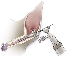
Figure 10-2 Smears of horizontal ear canal secretions may be collected by passing a cotton-tipped swab through the cone of an otoscope after otoscopic examination.
To prepare slides for routine staining, the swab is gently rolled onto a clean, dry slide in a thin layer, as thick smears are difficult to evaluate. Heat fixing neither systematically increases nor decreases numbers of yeast on specimens; although it is recommended by many to prevent loss of high lipid content, it is not necessary.3,10,11 After the material on the slide is allowed to air-dry, it is stained with any of the usual hematologic stains (e.g., Diff-Quik or Wright stain). It is recommended to have two sets of staining jars, one reserved for ear cytology and one reserved for other samples (e.g., blood smears, mass aspirates) as yeast and bacteria from ear cytologies may overgrow in the stain solution and contaminate other slides.
Cytologic Examination
Cerumen
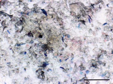
Cerumen, with its high lipid content, does not take up stain and provides the background for many normal ear swab cytologies (Figure 10-3).
Keratinocytes
Keratinocytes, including occasional nucleated forms, are noted in normal ears of both dogs and cats. Normal dogs were noted to have 3.9 keratinocytes per 40× high-power field (hpf) and normal cats were noted to have 8 per 40× hpf.13 The finding of nucleated forms should not be mistaken for a pathologic process (parakaratotic hyperkeratosis).
Bacteria
The ear canals of clinically normal dogs often contain small numbers of bacteria. The bacterial concentration typically is low enough that one sees only occasional or no bacteria on cytologic preparations (Figure 10-4). Many of these bacteria are potentially pathogenic and may colonize the ear canal when normal conditions are altered.3,6,8,14 In animals with bacterial otitis, cytologic evaluation of ear canal secretions often reveals large numbers of bacteria free in the smear (Figure 10-5). Unfortunately, no definitive rule exists for deciding if the bacteria are clinically relevant and warrant treatment. The decision should be based on severity of clinical signs and cytologic findings. Semiquantitative criteria to assess relevance of bacterial populations have been proposed on the basis of their numbers per 40× hpf as follows (Table 10-1): Bacterial counts expected in normal dogs vary among studies and have been reported as a median of 0 cocci (to averaging 5 cocci or fewer).14,12,13 Abnormal numbers have been reported to be an average of 25 or more organisms, with 6 to 24 organisms being in the “gray zone.” Bacterial counts expected in normal cats vary among studies and have been reported as a median of 0.3 cocci per 40× hpf in one study13 and an average of 4 or fewer cocci in a second study.12 Abnormal numbers have been reported to be 15 or more organisms, with 5 to 14 organisms being in the “gray zone.” Neither study identified bacterial rods as part of the normal ear cytology of dogs or cats.
TABLE 10-1
Malassezia and Bacteria: Expected Quantities
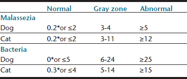
Proposed semiquantitative criteria for assessing organisms present in otic cytology based on median number (∗)per 40× hpf; or average numbers of organisms per 40× hpf.
Tater KC, Scott DW, Miller jr WH, Erb HN: The cytology of the external ear canal in the normal dog and cat, J Vet Med 50:370-374, 2003.
Ginel PJ, Lucena R, Rodriguez JC, Ortega J: A semiquantitative cytological evaluation of normal and pathological samples from the external ear canal of dogs and cats, Vet Dermatol 13: 151-156, 2002.
Angus JC: Otic cytology in health and disease, Vet Clin North Am Small Anim Pract 34:411-424, 2004.
Stay updated, free articles. Join our Telegram channel

Full access? Get Clinical Tree


