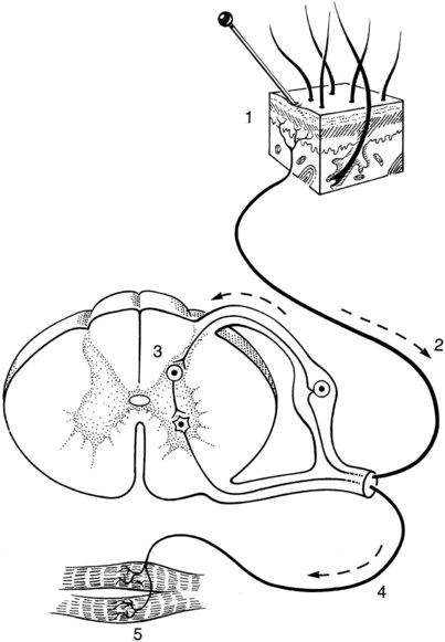
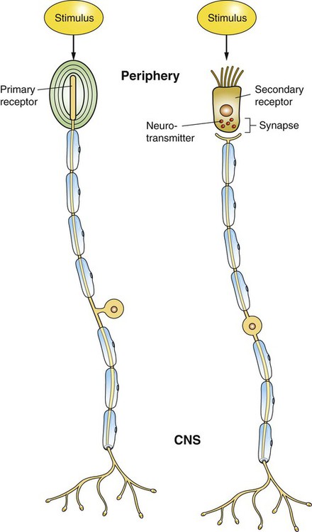
All reflex arcs contain five basic components (Figure 7-1). If any one of these five components malfunctions, the reflex response is altered. 1. All reflex arcs begin with a sensory receptor. Sensory receptors vary widely within the body but share a common function: they transduce a range of environmental energy, or the presence of an environmental chemical, into a cellular response that directly or indirectly produces action potentials along a sensory neuron. In other words, these receptors collect environmental signals and turn them into a format that can be understood by the nervous system. For example, receptors of the retina transduce light; those in the skin transduce heat, cold, pressure, and other cutaneous stimuli; muscle spindle receptors transduce stretch; and taste receptors transduce chemical stimuli from ingested material. A primary sensory receptor is a neuron with a specialized region for stimulus transduction (Figure 7-2; also see Figure 14-6, retinal photoreceptors). A secondary sensory receptor is a nonneural cell specialized for stimulus transduction that in turn affects neural activity by releasing neurotransmitter onto a neuron (see Figure 7-2; also see Figure 11-2, vestibular hair cells). Action potentials resulting from stimulus transduction are generated along sensory neurons at a frequency proportional to the intensity of the transduced stimulus. This proportionality between the intensity with which the receptor is stimulated and the frequency of the resulting sensory neuron action potentials is called frequency coding; it is one major way the receptor communicates to the central nervous system (CNS) the intensity of light, heat, stretch, and so forth, that it has transduced. Stronger stimuli will also activate a larger number of sensory receptors, known as the population code of stimulus intensity. 2. The next component in a reflex arc, alluded to earlier, is a sensory neuron (CNS afferent). These neurons carry action potentials, resulting from receptor activation, to the CNS. Again, in some cases the receptor is just a specialized, usually peripheral, region of the sensory neuron (primary receptors). In other cases the receptor is physically separate from and synapses on the sensory neuron (secondary receptors). Sensory neurons enter the spinal cord by way of the dorsal roots or enter the brain through cranial nerves. 3. The third component of a reflex arc is a synapse in the CNS. Actually, for most reflex arcs, more than one synapse occurs in series (polysynaptic). However, some reflex arcs that originate from the muscle spindle are monosynaptic. In polysynaptic reflexes, where one or more neurons lie between the sensory neuron input to the CNS and the motor neuron output, these interposed neurons are called interneurons and can be considered part of this third component of the reflex arc. 4. The fourth component is a motor neuron (CNS efferent), which carries action potentials from the CNS toward the synapse with the target (effector) organ. Motor neurons leave the spinal cord through the ventral roots, and motor neurons leave the brain through the cranial nerves. 5. The last component is some target organ (effector organ) that causes the reflex response. This is usually a muscle, such as the skeletal muscle fibers of the quadriceps muscle of the leg, in the case of the “knee jerk” (muscle stretch) reflex, or the smooth muscle of the iris in the pupillary light reflex. The target could also be a gland, such as a salivary gland in the salivary reflex. In reality, the final reflex response to a stimulus in mammals is rarely, if ever, the product of a monosynaptic reflex arc acting in isolation. Even if a sensory neuron participates in a monosynaptic reflex arc, it will often give off branches in the CNS that participate in polysynaptic reflex circuits. In addition, even the simplest mammalian reflex responses often involve both the excitation of a given muscle or muscles and the inhibition of another (usually antagonistic) muscle or muscles. The knee jerk reflex is a good illustration of these points (see Figure 8-3). With regard to the individual sensory neurons that underlie this reflex, some terminal branches make excitatory monosynaptic connections with motor neurons that activate the quadriceps muscle. Other terminal branches of that same sensory neuron participate in a disynaptic circuit that inhibits motor neurons innervating the antagonistic hamstring muscle.
The Concept of a Reflex
A Reflex Arc Contains Five Fundamental Components
![]()
Stay updated, free articles. Join our Telegram channel

Full access? Get Clinical Tree


