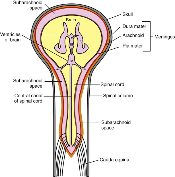
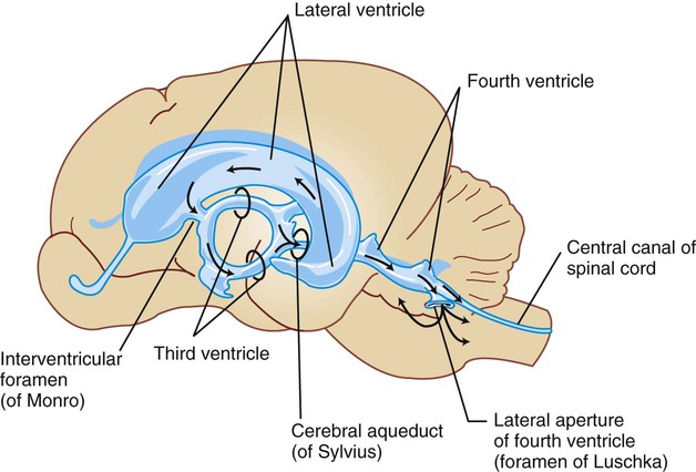
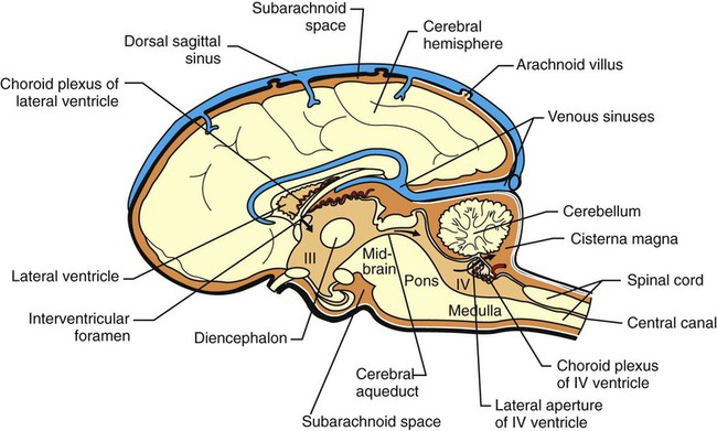
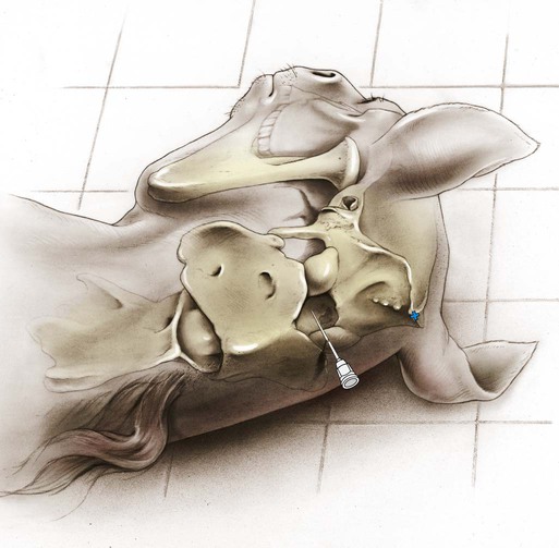
1. Cerebrospinal fluid has many functions. 2. Most cerebrospinal fluid is formed at the choroid plexus of the ventricles. 3. Cerebrospinal fluid flows down a pressure gradient through the ventricular system into the subarachnoid space. 4. Cerebrospinal fluid is absorbed into the venous system. 5. Hydrocephalus is an increased volume of cerebrospinal fluid in the skull. Cerebrospinal fluid (CSF) is a clear fluid present in the ventricles (core cavities) of the brain, in the central canal that runs through the core of the spinal cord, and in the subarachnoid space that surrounds the entire outer surface of the brain and spinal cord (Figure 15-1). The CSF contains almost no blood cells, little protein, and differs from plasma with respect to the concentration of various ions. Its rates of formation, flow, and absorption are sufficiently high to cause its replacement several times daily. Sampling its pressure, cell count, and levels of various biochemical constituents is a common diagnostic procedure for central nervous system pathology called a spinal tap. Injecting radiopaque dyes into the CSF of the subarachnoid space is the basis of a common neuroradiographic technique called myelography, often used in conjunction with computerized tomography (CT scan), which can assess the integrity of the spinal canal. Obstruction of the flow of CSF produces a condition called hydrocephalus. An understanding of the formation, flow, and absorption of CSF is essential for understanding these diagnostic procedures and the pathophysiology of hydrocephalus. The ventricles are a series of interconnected cavities in the core of the brain that have an ependymal cell lining and are filled with CSF (Figure 15-2). The lateral ventricles are respectively located in the two cerebral hemispheres, the third ventricle is found at the midline of the diencephalon, and the fourth ventricle is located between the cerebellum and the dorsal surface of the hindbrain (pons and medulla) (Figure 15-3). The majority of CSF is formed by a choroid plexus located in each of the four ventricles. These are small, cauliflower-like growths of clustered villi that form a portion of the floor or roof of each ventricle (see Figure 15-3). The plexuses consist of tufts of capillaries covered by a layer of ependymal cells. These ependymal cells, unlike the cells lining the rest of the ventricle, form a selective, tight-junction barrier to the secretions of the leaky capillaries and to other surrounding fluids (e.g., CSF, extracellular fluid). Membrane transporters and selective channels regulate the passage of ions and molecules across the ependymal cell barrier, effectively controlling the composition of the CSF being synthesized in the ventricle. The active transport of sodium ions (Na+) contributes to a net movement of sodium chloride (NaCl) into the ventricles. This osmotic gradient regulates the water content of the CSF as water follows the NaCl passively into the ventricle. There is evidence that some potentially harmful metabolic waste products deposited in the CSF can actually be absorbed and removed by the choroid plexus. CSF flows, by bulk, down a pressure gradient from its site of formation at the choroid plexuses through the ventricular system and subarachnoid space into the venous system. Fluid formed in the lateral ventricles passes into the third ventricle through the interventricular foramina (foramina of Monro) (see Figures 15-1, 15-2, and 15-3). The fluid mixes with fluid formed in the third ventricle and from there passes through the cerebral aqueduct (aqueduct of Sylvius) of the midbrain into the fourth ventricle. Fluid in the fourth ventricle passes into the subarachnoid space through two lateral apertures or foramina of Luschka. Some mammals have a third, medially located passageway from fourth ventricle to subarachnoid space (foramen of Magendie). Recall that the brain and spinal cord are encased in bone (the skull and spinal canal, respectively) and are covered by a series of three membranes called the meninges (see Chapter 3). From outer to inner, these membranes are the dura, arachnoid, and pia (see Figure 15-1). The subarachnoid space lies between the arachnoid and pia, and when the CSF exits the brain through the apertures (foramina) of the fourth ventricle, the CSF fills the subarachnoid space, spreading out over the entire outer surface of the brain and spinal cord. Thus the entire CNS is essentially floating in a fluid-filled, membranous bag. As the CSF circulates up over the dorsal convexity of the brain, it is absorbed into the venous system near the midline. The pressure, cell count, and chemical constituents of CSF can be sampled by placing a styletted spinal needle into the subarachnoid space. Anatomically, the most convenient place to perform this varies with species. In humans it is usually performed in the lumbar spinal column because the human spinal cord tapers to a cone (conus medullaris) near the first lumbar vertebra (humans have five lumbar vertebrae) as the dura and arachnoid continue down to around the second sacral vertebra. This provides a relatively large subarachnoid space (lumbar cistern) in the human midlumbar spinal column from which to sample. In most veterinary species, however, the conus medullaris extends to about the sixth or seventh lumbar vertebra, leaving only a small subarachnoid space in the spinal column. Therefore, most veterinary spinal taps are performed by sampling from the subarachnoid space region that is accessed between the skull and the first cervical vertebra (atlas) in anesthetized animals (Figure 15-4). In this location the subarachnoid space, formed as the arachnoid stretches from the caudal cerebellar surface to the dorsal surface of the medulla, is called the cisterna magna (“big cistern”; also called cerebellomedullary cistern) and is much deeper than other portions of the subarachnoid space (see Figure 15-3). Spinal taps provide valuable information about such neuropathological lesions as intracranial space-occupying masses and inflammation.
Cerebrospinal Fluid and the Blood-Brain Barrier
Most Cerebrospinal Fluid Is Formed at the Choroid Plexus of the Ventricles
Cerebrospinal Fluid Flows Down a Pressure Gradient Through the Ventricular System Into the Subarachnoid Space
< div class='tao-gold-member'>
![]()
Stay updated, free articles. Join our Telegram channel

Full access? Get Clinical Tree


Cerebrospinal Fluid and the Blood-Brain Barrier
Only gold members can continue reading. Log In or Register to continue
