Tachypnea and Hypoxemia

Chapter 9 Tachypnea and Hypoxemia


TACHYPNEA WITH HYPOXEMIA


When the tachypneic patient is determined to be also hypoxemic, the diagnostic and therapeutic approach will need to be tailored appropriately. Respiratory distress in small animals often presents a therapeutic dilemma. Tachypneic hypoxemic patients can be so compromised that diagnostic tests can stress them to the point of respiratory and cardiac arrest. A diagnosis should not come at the expense of the patient. Restraint for catheterization, radiographs, and physical examination may have to wait until the patient is relaxed and breathing more easily.


When an animal has difficulty oxygenating blood, breathing becomes more labored. Although terms such as dyspnea or anxiety should be avoided in veterinary medicine (our patients cannot tell us they’re having difficulty breathing or fear), we should assume that these patients are experiencing tachypnea and orthopnea associated with hypoxemia. This natural response leads to more complications because the stressed patient needs more oxygen and rapid breathing may not be as efficient as relaxed, normal breathing. For this reason, tranquilizers and sedative drugs may prove very useful in the early treatment of respiratory distress.


Inspired oxygen concentration may be increased using a face mask, nasal cannula, or an induction chamber attached to an anesthesia circuit’s oxygen supply. Oxygen cages increase inspired oxygen while allowing the clinician time to observe the patient and localize the problem.


It is important to observe the patient, refine the list of differential diagnoses, and determine the nature of the problem. A rapid, shallow (restrictive) respiratory pattern suggests higher than normal elastic forces within the lung. A deep, noisy (inspiratory) pattern is seen with increased airway resistance from airway obstruction. With a restrictive pattern, auscultation can help differentiate pleural space disease (pneumothorax, hydrothorax) from parenchymal diseases (pneumonia, pulmonary edema). Signalment and history can help determine a cause of airway obstruction (brachycephalic airway disease, history of a cough, playing with small toys).2-4


A basic understanding of the work of breathing and mechanisms of impaired gas exchange will help the clinician develop a rational diagnostic and treatment plan.


WORK OF BREATHING


For normal respiration and gas exchange, fresh gas must be brought into the alveoli through the process of alveolar ventilation. The oxygen cost of quiet breathing is extremely small, less than 3% of total resting oxygen consumption. With voluntary hyperventilation (as is seen with respiratorydistress), it is possible to increase cost of breathing to 30% of resting oxygen consumption. For animals with obstructive or restrictive lung disease, oxygen cost of breathing can limit their exercise tolerance.1,5


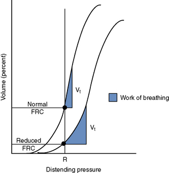
Muscles of the respiratory system must overcome two major forces in normal respiration: elastic recoil and airway resistance. The functional residual capacity (FRC) is defined as the volume of air remaining in the lungs after a normal expiration. FRC is the volume of the lungs at rest and is determined by the static properties of the respiratory system.1,5 Fibrous structures of the lung provide elastic recoil. The fibrous structures of the lung favor pulmonary collapse, while the fibrous structures of the chest wall favor thoracic expansion. The balance between lung elastic recoil and thoracic wall recoil ultimately determines the FRC. The FRC is reduced in patients with reduced lung compliance.1,5


To understand how FRC is clinically significant we must understand compliance. Pulmonary compliance is reflected by the slope of the pressure-volume curve (Figure 9-1). The slope of this curve represents the volume change per unit pressure.1,5 When the slope of this curve is steep, a small change in pressure results in a large change in volume. This makes for efficient ventilation requiring minimal energy to effect gas movement. Diseases affecting pulmonary compliance can increase the work required for normal ventilation. Pulmonary atelectasis, pneumonia, pulmonary edema, fibrosis, and pleural space diseases (causing pulmonary collapse) are examples of conditions resulting in decreased pulmonary compliance.


Stay updated, free articles. Join our Telegram channel

Sep 10, 2016 | Posted by in SMALL ANIMAL | Comments Off on Tachypnea and Hypoxemia

Full access? Get Clinical Tree

Get Clinical Tree app for offline access