Surgical Repair of Vaginal Anomalies in the Bitch

Chapter 213


Surgical Repair of Vaginal Anomalies in the Bitch


A number of developmental and acquired conditions affect the canine vagina and vulva. Congenital conditions, which affect primarily younger dogs, include rectovaginal fistula; vulvar/vaginal hypoplasia; anovulvar cleft; clitoral enlargement; and vaginal septa, bands, or stenosis. Acquired conditions generally affect older dogs and include vaginal neoplasia and vaginal prolapse. Some acquired conditions such as vaginal hyperplasia and perivulvar dermatitis can affect younger dogs. Invariably congenital and acquired abnormalities overlap somewhat because some acquired anatomic problems may develop secondary to congenital abnormalities within the reproductive tract. Surgery to correct these abnormalities often corrects the presenting clinical signs. These procedures may be as simple as digital breakdown of a thin vaginal band or more complex (e.g., complete vaginal ablation).


Surgical Approaches to the Canine Vagina


Most surgical procedures of the vagina (and vestibule) can be approached via a perineal or caudal episiotomy. More involved procedures may require a ventral approach, which may necessitate a combined abdominal approach with or without a pubic osteotomy. With either surgical approach, strong consideration should be given to management of postoperative pain. The use of a fentanyl patch or epidural analgesia should be considered before surgery. An appropriately sized fentanyl patch (4 µg/kg/hr in dogs) should be applied the day before surgery. Alternatively, an epidural with the use of preservative-free morphine (0.1 mg/kg), bupivacaine (0.5 to 1 mg/kg), or a combination of both can be administered before or immediately after surgery. If epidural analgesia is elected, the surgeon should review the various techniques and dosing guidelines described in the veterinary literature.


Caudal Approach with Episiotomy


When performing an episiotomy, the surgeon should place the animal in sternal recumbency with the pelvic limbs hanging over the end of the table. The edges of the table should be well padded to avoid trauma to the limbs. The table is tilted so that the animal’s head is down about 30 degrees and the vulva is at a comfortable working height. This head-down position may make ventilation of the patient more difficult, requiring assisted manual or mechanical ventilation to maintain adequate oxygenation and anesthetic plane. These patients are also at increased risk of gastric reflux/regurgitation and subsequent aspiration. These animals must be fasted at least 12 hours before surgery and receive H2 blockers to decrease gastric acidity. A loading dose of metoclopramide (1 mg/kg IV), followed by a constant rate infusion (1 mg/kg/hr) during anesthesia also may reduce the incidence of gastroesophageal reflux. In addition, a cuffed endotracheal tube should be used, and the pharynx should be evaluated and suctioned at the end of the procedure. A purse-string suture is placed around the anus to prevent fecal contamination of the surgical field. A piece of surgical tape marked “purse string” should be placed on the patient’s head to remind the surgeon and anesthetist to remove the sutures once the procedure is finished. The vestibule and caudal vagina is flushed with dilute povidone-iodine solution as part of the surgical scrub. Three to four flushes of a 1 : 10 dilution of povidone-iodine with sterile water should be used to minimize vaginal mucosal irritation.


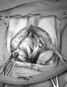
An incision along the median raphe is made from the level of the caudodorsal aspect of the horizontal vaginal canal, descending to the dorsal commissure of the vulvar cleft. The incision is continued along the same plane of the skin incision through the vaginal musculature and mucosal layers. Placement of a flat instrument (e.g., scalpel handle) in the vestibule can be used to stabilize the tissues while the incision is made through the dorsal vestibular mucosa. Alternatively, Metzenbaum scissors can be used to cut the mucosal layer. Cautery, vessel ligation or compression of the vestibular wall using two Doyen bowel clamps (positioning one on each side with one blade in the vestibular lumen and one on the skin surface) can be used for hemostasis (Figure 213-1). Hemorrhage often is associated with surgery to this region because of the increased vascularity to the vaginal tissues. Exposure is maintained with the use of self-retaining retractors (i.e., Gelpi, Weitlander, or ring retractors) or stay sutures. A urinary catheter should be placed if there is potential for tissue manipulation around the urethra and urethral tubercle.


Closure of the episiotomy is performed in four layers: mucosa, muscular tissue, subcutaneous tissue, and skin. A simple interrupted or continuous pattern of 3-0 monofilament absorbable suture is used for the mucosa. The muscular and subcutaneous tissues can be closed together or separately, depending on the size of the animal, using a simple continuous pattern of 3-0 or 4-0 absorbable suture. The skin edges can be closed with sutures (simple interrupted or cruciates) or surgical staples.


Perineal Approach with Episiotomy


In some cases, the episiotomy can be modified by limiting the approach to the perineum. This approach allows access to the vestibulovaginal region without incising into the dorsal aspect of the vestibule, therefore limiting the intraoperative hemorrhage and minimizing postoperative pain. It can be used for resection and anastomosis of vestibulovaginal stenosis and to access pedunculated masses of the vagina or vestibule. This approach also can be combined with a ventral abdominal approach for vaginal ablation.


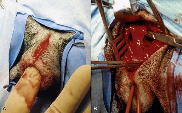
An incision along the median raphe is made from the level of the caudodorsal aspect of the horizontal vaginal canal, descending to the dorsal commissure of the vulvar cleft. Digital palpation during the approach facilitates identification of the vagina and helps identify the location of interest (Figure 213-2, A). The incision is continued along the same plane of the skin incision until the muscular wall of the vagina/vestibule is reached. Cautery vessel ligation should be used for hemostasis. Exposure is maintained with the use of self-retaining retractors. Blunt dissection can be performed around the vagina as needed (Figure 213-2, B). Careful dissection along the ventral aspect of the vagina is necessary to avoid trauma to the urethra. A urinary catheter should be placed if there is potential for tissue manipulation around the urethra and urethral tubercle.


Closure of the perineum is performed in two layers: subcutaneous tissues and skin. The subcutaneous tissues can be closed together or separately, depending on the size of the animal, using a simple continuous pattern of 3-0 or 4-0 absorbable suture. The skin edges can be closed with sutures (simple interrupted or cruciates) or surgical staples.


Ventral Approach


Fortunately, the ventral approach to the canine vagina is not used often because it requires a pelvic osteotomy. The ventral approach often is used for vaginal ablations. However, surgery through an abdominal or caudal/perineal approach often is possible to avoid the need for a pelvic osteotomy (see next section). The urethra should be catheterized to aid in identification and prevent iatrogenic trauma.


A standard ventral midline approach is performed in the caudal abdomen up to the cranial pelvic rim. The urinary bladder can be manipulated aside to provide access to the vagina. The pelvic osteotomy is performed to increase the exposure to the vagina and urethra. The incision is extended caudally over the midline of the pubic symphysis. The adductor muscles are elevated laterally with periosteal elevators to expose the pubic and ischial rami. A partial or complete pelvic osteotomy can be performed, depending on the location and amount of exposure needed. A partial pelvic osteotomy involves osteotomies through the pubic rami and across the pubic symphysis at the level of the obturator foramen. A complete pelvic osteotomy through both pubic rami and ischial rami gives the greatest exposure. Once the surgeon has determined the location of the osteotomy, holes are predrilled on either side of the osteotomy site. It is much easier to drill these holes before performing the osteotomy, especially if using a hand chuck and pin. The osteotomy is performed with Gigli wire, rotating burr, sagittal saw, or bone cutters. The internal obturator muscle is elevated off the pubic symphysis on one side of the pelvis and hinged on the contralateral internal obturator muscle to expose the pelvic canal.


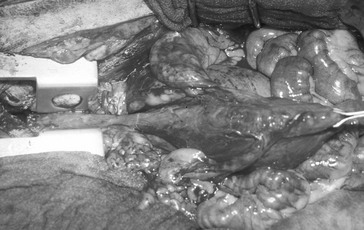
Alternatively an osteotomy through the pubic symphysis, separating the hemipelvis, can be performed. The flexibility of the pelvis allows the placement of retractors to separate the hemipelvis, giving exposure to the pelvic canal. Holes can be predrilled on either side of the pubic symphysis. Self-retaining retractors (i.e., Finochietto retractors) facilitate exposure (Figure 213-3). Care must be taken not to put excessive stress on the hemipelvis, which can create a fracture or sacroiliac luxation (especially in young dogs or cats). Closure of this approach begins with reduction of the pelvic floor, using 18- to 20-gauge cerclage wire. The predrilled holes allow for rapid and accurate alignment of the pubic and ischial rami. The obturator and adductor muscle fascia from either side is sutured to its contralateral partner along the midline. Closure of the linea, subcutaneous tissue, and skin is performed routinely. Because of the osteotomy, activity should be restricted for 4 months after surgery. In small patients the pubic symphysis does not have to be replaced. Closure of the obturator and adductor muscles along the midline provides adequate support of the pelvic floor.


Combined Abdominal and Caudal/Perineal Approach


In most cases, the cranial aspect of the vagina can be accessed through a caudal abdominal approach combined with a caudal or perineal approach, avoiding the need for a pubic osteotomy. This approach can be used for vaginal ablation secondary to a vaginal neoplasia and can be combined with an ovariohysterectomy if the patient is intact.


Once the caudal abdomen is approached via a routine midline laparotomy, the bladder can be retroflexed to allow exposure to the vagina and associated structures. Fascial and peritoneal attachments between the vagina and rectum are bluntly dissected free. Similarly, dissection of attachments between the vagina and the urethra is performed, avoiding any disruption of the craniolateral aspect of the urethra and the periurethral tissues and avoiding damage to the ureters and urethral innervations. The cranial and caudal branches of the vaginal artery and vein are ligated. Once the vagina is dissected free, a stay suture can be placed through all layers of the vagina and the loop of the suture pushed caudally or passed into the vaginal lumen. The laparotomy is closed routinely.


The intraluminal stay suture then can be grasped via a caudal approach with episiotomy; the extraluminal stay suture can be grasped via a perineal approach. Once the stay suture is identified, it is retracted caudally, withdrawing the cranial vagina into the perineal site, allowing for complete resection of the vagina.

< div class='tao-gold-member'>

Stay updated, free articles. Join our Telegram channel

Jul 18, 2016 | Posted by in PHARMACOLOGY, TOXICOLOGY & THERAPEUTICS | Comments Off on Surgical Repair of Vaginal Anomalies in the Bitch

Full access? Get Clinical Tree

Get Clinical Tree app for offline access