Surgical procedures for the conjunctiva and the nictitating membrane

Chapter 7 Surgical procedures for the conjunctiva and the nictitating membrane


Introduction


Conjunctival and nictitating membrane diseases occur frequently in all animals. In the dog, the majority of conjunctival inflammations are secondary, and bacteria isolated from these eyes usually yield a variety of normal residents of the conjunctival surfaces. In the cat, most conjunctival inflammations are primary, infectious, and associated with viruses, chlamydia, and mycoplasma. In horses, primary conjunctivitis is infrequent, but both the conjunctiva and the nictitans are often involved with neoplasia in older horses. In cattle, primary inflammations and neoplasia of the conjunctiva and nictitans are frequent.


Surgery for conjunctival diseases in the dog and cat is infrequent except for excision of conjunctival masses, which are often combined with other surgical procedures involving the eyelids. The most frequent conjunctival surgical procedures are the several different types of conjunctival grafts or flaps for treatment of corneal ulcers that threaten the integrity of the globe, and the maintenance of vision. Fortunately, there is ample and mobile conjunctiva for the construction of these grafts. The success rates for the different conjunctival autografts are very high (>90%), and their value in the overall clinical management of small animal patients is often understated. In horses and cattle, neoplasia is often treated with surgery, sometimes combined with cryotherapy or local chemotherapy.


The nictitating membrane has several synonyms including membrana nictitans, palpebra tertia, plica semilunaris, third eyelid, and the haws. The nictitating membrane is a semilunar fold of conjunctiva, which also occurs in redimentary form in humans and non-human primates, that protrudes from the medial canthus and can extend over a significant portion of the cornea. It is more mobile in birds and cats, and in birds it is very thin and semitransparent.


Diseases of the nictitating membrane are not infrequent in small animals. In dogs and cats, congenital, inflammatory, traumatic, and neoplastic disorders may be treated surgically. In cats, more frequently than in dogs, disorders of the nictitating membranes may also signal serious systemic diseases. In both horses and cattle, neoplasms are the most frequent surgical indications.


Movement of the nictitating membrane somewhat parallels its development in the different species. In dogs, the dorsolateral movement of the nictitating membrane appears passive and associated with movement of the retractor oculi muscle, and the shifting of the endorbita fascia and orbit fat pad. As the globe is retracted into the depths of the orbit, the orbital adipose tissues shift forward to push the base of the nictitating membrane anteriorly and protract the structure across the cornea. Fascial attachments in the canine nictitating membrane attach the base of the hyaline cartilage and the nictitans tear gland to the bulbar fascia, medial canthal ligament, and endorbita or periorbita fascia.


In cats, movements of the nictitating membrane result from contraction of smooth muscles, and are associated with its sympathetic nerve supply. Stimulation of the preganglionic cervical sympathetic nerve produces frequency-dependent contractions of the nictitating membrane. The feline nictitating membrane contains two thin sheets of muscle, named by Acheson in 1938 as the medial and inferior muscles (also called Müller’s muscles), that arise deeply within the orbit from the periorbital fascia covering the medial and ventral rectus muscles to insert into the adjacent sides of the T-shaped nictitans cartilage. Alpha1-adrenoceptor antagonists, whether selective or non-selective, produce a dose-dependent depression of the evoked nictitating membrane movements, suggesting that the feline nictitans contains mainly α1-adrenoceptors postsynaptically.


In horses and cattle, movements of the nictitans result from contraction of the rectus and retractor oculi muscles which displace the orbital fat rostrally and protract the nictitans across the cornea in a more horizontal direction.


In birds, the active movement of the nictitating membrane is highly developed, and is rapidly protracted over the cornea toward the lateral canthus by the pyramidalis muscle. Protraction of the third eyelid is accomplished by contraction of the striated pyramidalis muscle that originates in the posterior sclera and attaches to the nasal edge of the nictitans. The quadratus muscle forms a sleeve dorsal to the optic nerve through which the tendon of the pyramidalis muscle passes, allowing a pulley action of the quadratus muscle to amplify or modify action of the pyramidalis. Innervation of both muscles by the sixth cranial nerve is widely cited. The leading edge is pigmented and forms a marginal plait (plica marginalis). The avian nictitans can be very thin and difficult to appose by sutures after trauma.


Conjunctival anatomy


Conjunctival anatomy is similar in all mammals. The conjunctiva spans the eyelid margins to the limbus of the globe and is the major barrier to the external environment for the globe and orbit. The conjunctiva, as a mucous membrane, forms the first line of defense against the external elements. Its elastic nature accommodates both globe and eyelid movements. The conjunctiva rests on the endorbita or periorbita fascia; on the globe the conjunctiva is in intimate contact with the bulbar fascia (or Tenon’s capsule) and attaches at the limbus. As a result, the conjunctiva accommodates globe mobility and provides for nearly unrestricted ocular movements. The conjunctiva lining the inner aspects of the eyelids and fornix for 360° is next to the fibrous tarsus, septum orbitale, and the endorbita. The collective conjunctiva is often referred to as the conjunctival sac.


Traditionally the conjunctiva is divided macroscopically into palpebral, fornix, and bulbar components. The palpebral conjunctiva lines the inner aspects of the eyelids. The bulbar conjunctiva mucosa starts at the corneal epithelial layer at the limbus. It covers Tenon’s capsule or bulbar fascia, and extends to join the palpebral conjunctiva at the fornix or the conjunctival cul-de-sac. Tenon’s capsule or bulbar fascia is quite thin in cats, horses, and cattle but variable in dogs, sometimes 5 mm or more thick. Medially a conjunctival apex or fold, the nictitating membrane or third eyelid, divides the ventral conjunctival fornix into two parts: the outer or palpebral portion, and the deeper or bulbar component (Fig. 7.1). As the conjunctival fornices span the globe and eyelids for 360°, the fornices may be divided into dorsal, lateral canthal, ventral, and medial canthal parts. The dorsal conjunctival fornix is deeper than the ventral fornix, and this may be necessary to accommodate downward eye movements. The ventral conjunctival fornix is shallower and its primary function is as a collecting basin for the tears. The medial conjunctival fornix is divided into anterior and posterior fornices of the nictitating membrane. Medial movement of tears toward the upper and, more importantly, the lower lacrimal puncta by the eyelid is not understood, but it appears to be an active rather than a passive process.


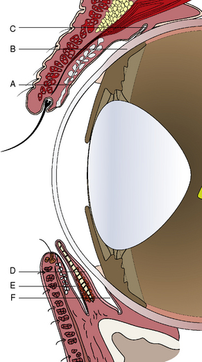
The palpebral conjunctiva originates at the eyelid margin (the margo-intermarginalis) and the orifices of the meibomian glands. The margo-intermarginalis is the last tissue that when contact occurs with the cornea and conjunctiva produces no damage; when the outer leading edge of the eyelid skin touches the cornea, as in entropion, corneal and conjunctival damage start! The palpebral conjunctival surface epithelium at the eyelid margin consists of non-keratinized stratified epithelium, but changes after several millimeters from the lid margin into pseudostratified columnar epithelium. Immediately beneath the palpebral conjunctiva is the thicker connective tissue, the tarsal layer, which contains the sebum-producing tarsal or meibomian glands. Once the pseudostratified epithelial surface is established, conjunctival goblet cells begin to appear and are most numerous in the fornices. These goblet cells produce mucin, an important deep component of the preocular or precorneal film, and an essential lubricant to prevent eyelid trauma to the conjunctival and the corneal surfaces (Fig. 7.2). Mucin forms the innermost layer of the preocular (precorneal) film and ranges in thickness from 1 μm on the cornea to 2 μm or more on the conjunctival surfaces. Mucin, a hydrated glycoprotein, forms an interface between the larger aqueous portion of the preocular film and the hydrophobic corneal epithelium. A relatively small portion of mucin is water soluble and part of the middle or aqueous fraction of the preocular film. The goblet cell-derived mucin decreases the surface tension of the preocular film, enhances the stability of the preocular film, and aids in the coherence of the aqueous portion of the preocular film to the corneal and conjunctival epithelia. Mucin also coats and reduces the irregularities of the corneal epithelium to produce an optically smooth corneal surface. The palpebral conjunctiva is quite transparent and often allows visualization of impacted or inflamed meibomian (or Meibom) glands on the deep aspects of both eyelids.


The bulbar conjunctiva consists of entirely pseudostratified epithelium, and is most firmly attached at the fornices and the limbus. About 3 mm from the corneal periphery, the bulbar conjunctiva, Tenon’s capsule (orbital fascia), and the limbus become closely united. Because of its surface area and availability, the dorsal bulbar conjunctiva is the principal source of mucous membrane for graft construction and transplantation to the cornea. The bulbar conjunctiva contains few goblet cells.


The limbal conjunctival cell may have special significance as a critical stem cell for conjunctival surgical procedures. Transplantation of limbal stem autografts to defects in the corneal epithelium in humans successfully re-establishes the corneal epithelial characteristics and clarity, and offers potential for animals. Normal corneal epithelial cells migrate centripetally from the periphery to the center of the cornea. The limbal cells appear to be the corneal epithelial stem cells and capable of additional cell multiplication and differentiation, and a terminal transparent cell. Epithelial cells from the limbus readily grow as explants; however, peripheral and central corneal explants are progressively less likely to grow as explants. The peripheral and, to a greater extent, the central corneal epithelial cells appear committed to terminal differentiation. As a result, both the limbal and conjunctival epithelia are excellent sources for autografts.


Lymphoid follicles are scattered throughout the conjunctiva, but may be more numerous in the fornices. These lymphoid follicles, acting as regional lymph nodes, are the major defense for the conjunctival surfaces, and during inflammation increase both in size and number. Grossly lymphoid follicles appear as clear-to-translucent raised circular areas.


The substantia propria of the conjunctiva consists of two layers: a superficial layer that contains lymphatic follicles and glands, and the deeper fibrous layer that attaches the conjunctiva to the orbital and eyelid fascia. The latter layer tends to be quite variable in dogs, and in certain breeds is sometimes quite thick; however, in contrast, it tends to be quite thin in cats, horses, and cows. In the preparation of bulbar conjunctival grafts, the thickness of this layer can contribute to excessive tension of the graft and retraction toward the limbus. The nerves and vessels of the conjunctiva are primarily in the deep layer, and are derived from the anterior ciliary arteries that are branches from the external ophthalmic arteries. Additional arterial branches arise from the superficial temporal, malar, and palpebral arteries. Venous drainage from the conjunctiva occurs to adjacent palpebral and malar veins that eventually join the facial vein, or deep into the orbit with the superficial angularis oculi vein to the orbital plexus and superficial temporal vein. The lymphatics of the conjunctiva are divided into superficial and deep systems. Lymphatic drainage from the medial aspects of the conjunctiva is to the submaxillary lymph nodes and laterally to the parotid lymph nodes.


Anatomy of the nictitans


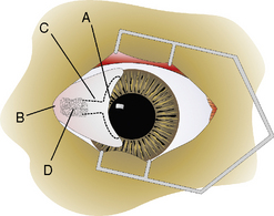
The gross anatomy of the nictitating membrane is quite similar among mammals. Located in the medial canthus, the nictitating membrane is a roughly triangular-shaped fold of conjunctiva, with the base of the triangle consisting of its free or leading margin (Fig. 7.3). Both anterior (palpebral) and posterior (bulbar) surfaces are confluent with the palpebral and bulbar conjunctival mucosa. Its free margin or border is usually pigmented in animals. When non-pigmented, the nictitans appears more prominent. Within the substance of the nictitating membrane is a hyaline T-shaped cartilage plate, which helps provide rigidity to the structure, assists conformation to the corneal curvature, and prevents disfigurement during movement (Fig. 7.4). The ‘arms’ of the T-shaped cartilage are immediately under its leading margin, and are relatively thin and slender compared to the thicker stem or base. The superficial gland of the nictitating membrane in both dogs and cats surrounds the base of the nictitans cartilage and produces seromucoid tears. Both dogs and cats possess a single nictitans gland, but in some species such as birds, the third eyelid gland may have two divisions. The deeper avian third eyelid gland is referred to as the Harderian gland.


In horses, the nictitans gland is quite large (19 mm anteroposterior × 11 mm wide). In cattle, the triangular-shaped nictitans gland (41 mm long × 26 mm wide) is in two parts, although it appears as one confluent structure: the anterior seromucoid nictitans gland which surrounds the cartilage shaft (serous-appearing acini, but periodic acid–Schiff positive), and the deeper Harderian gland, which also has two parts, and is also mucoid but periodic acid–Schiff negative.


The nictitans gland in small animals is an important accessory tear-producing gland providing about 25–40% of the total tears. Lymphoid follicles are usually present on the posterior or bulbar surface of the nictitans, appearing as raised translucent spots, and especially prominent in the horse. Lymphoid follicles are infrequent on the anterior or palpebral surface of the nictitating membrane and often signal chronic conjunctival irritation.


The blood supply to the canine nictitating membrane is derived from a branch of the internal maxillary artery located within the space between the ventral and medial rectus muscles. Smaller branches are often located on both sides of the stem portion of the cartilage and should be avoided during surgery. Sensation is provided by the infratrochlear nerve branch of the ophthalmic nerve, a subdivision of the trigeminal nerve.


The primary functions of the nictitating membrane are to assist in the protection of the cornea and provide the second largest portion of tears. Mucin from the third eyelid tears forms an essential part of the preocular film. Nictitating membrane movement may assist in the movement of tears to the medial canthus, and the ‘pick-up’ of the tears by the lacrimal puncta. Loss of the nictitans results in a larger medial lacrimal lake or conjunctival sac and often chronic conjunctivitis due to the collection and impaired drainage of tears from this area. The nictitating membrane is an essential component of the conjunctiva as well as the tear-producing system. Total excision of the nictitating membrane should be reserved for extensive neoplastic involvement of this structure.


Bulbar/palpebral conjunctival biopsy (punch/snip)


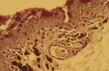
Biopsy of the conjunctiva may be indicated for diagnosis of non-specific diffuse and focal conjunctival inflammations (Fig. 7.5), and for possible neoplasia in all animal species. For biopsies of suspected conjunctival inflammations, selection of the ventral conjunctiva may be more rewarding. Focal swellings, such as conjunctival cysts, parasitic granulomas, nodular granulomatous episclerokeratitis, proliferative keratoconjunctivitis of Collies, and nodular fasciitis, can be biopsied or removed for histologic examination.


In contrast to the majority of eyelid neoplasms in the dog, primary conjunctival neoplasms may be invasive locally and should be widely excised. Hemangiomas, hemangiosarcomas, angiokeratomas, viral papillomas, squamous cell carcinomas, and malignant melanomas have been reported in the dog. In the cat, the most frequent conjunctival neoplasm is squamous cell carcinoma. Neoplasms of the conjunctiva affect the dorsal to dorsolateral limbal area most frequently, suggesting solar (ultraviolet) radiation may play an important role in their genesis. If a conjunctival neoplasm penetrates the anterior orbital fascia, intraorbital extension is likely and the clinical appearance of the neoplasm may be deceptive. In the clinical assessment before excision, involvement of the deeper layers of the conjunctiva should be determined by ultrasonography or other imaging procedures.


If the mass involves the superficial layers of the conjunctiva, it is usually easily manipulated and moves with the conjunctival mucosa. If the tumor has extended into the deeper submucosa or even infiltrated the periorbital fascia, the tumor usually remains fixed as the surrounding conjunctiva is manipulated.


In horses and cattle, solitary masses of the conjunctiva are assumed to be neoplastic until proven different histologically. In both species, squamous cell carcinomas are the most frequent neoplasms, affecting the eyelid margin, nictitans, and limbus. Because of malignancy and infiltration of adjacent tissues, surgery is often combined with other treatment modalities, such as cryotherapy.


Sedation or short-acting general anesthesia is usually necessary for conjunctival biopsy. Topical anesthesia, such as 0.5% proparacaine or 0.5% tetracaine, can also be used. The eyelids are not usually clipped or prepared for surgery. The conjunctival surfaces, including the fornices, are cleansed with sterile cotton-tipped applicators and 0.5% povidone–iodine solution.


The elastic and accessible conjunctiva can be easily biopsied. Small biopsies less than 1 cm do not require sutures and readily heal by secondary intention. The eyelids are retracted with a small wire speculum, conjunctival area to be biopsied is elevated by small thumb forceps, such as the Bishop–Harmon, and the conjunctiva excised by small tenotomy scissors. If the conjunctival defect is greater than 1 cm, the mucosa edges are apposed with 4-0 to 7-0 simple interrupted or continuous absorbable sutures. Postoperative treatment usually consists of topical antibiotics or antibiotics/corticosteroids administered three or four times daily for several days.


Surgical repair of conjunctival lacerations


Conjunctival lacerations are infrequent and usually combined with lacerations of the eyelids, cornea, and sclera. The presence of bulbar conjunctival lacerations signals the need for a complete eye examination. Bulbar conjunctival lacerations may mask more serious and vision-threatening full-thickness scleral lacerations and intraocular damage.


Small conjunctival lacerations less than 1 cm will usually heal by secondary intention. These lacerations should be carefully examined to exclude any intraocular damage, and any foreign material removed manually or irrigated from the tissues. Topical antibiotic solutions are administered several times daily for 5–7 days, or until the conjunctival epithelium has bridged the wound.


Larger conjunctival lacerations are usually apposed by sutures. Palpebral conjunctival wounds often involve the full-thickness eyelids. Large bulbar conjunctival lacerations should be approached with caution, as intraocular tissue involvement is likely. If hyphema is present with a bulbar conjunctival laceration, intraocular damage has occurred, and careful examination of the entire globe is warranted. Both direct and indirect trauma can result in intraocular hemorrhage and inflammation.


Large conjunctival lacerations (>1 cm) are best treated by apposition with sutures. After short-acting general anesthesia, the conjunctival surfaces are carefully cleaned with cotton-tipped applicators and, if necessary, with serrated thumb forceps. Any foreign material should be removed and, if possible, identified. Vegetative material will often provoke an acute and intense inflammation. Fungal organisms may also be introduced into the tissues. Debridement of conjunctival tissues, like that of the eyelids, should be minimal to preserve as much of the conjunctiva as possible. Small thumb forceps with 1 × 2 teeth and a small Castroviejo needle holder are used. Simple interrupted absorbable sutures (usually 5-0 to 7-0) are used for apposition.


Some chemosis is anticipated postoperatively. Topical antibiotics, often combined with corticosteroids, are instilled four to six times daily for 5–7 days. The ointment form of medication may be more advantageous as drug contact time is prolonged, and the ointment coats the conjunctival and suture surfaces to act as a lubricant. If the conjunctival swelling is excessive, hot and cold packs to the area may promote local circulation and reduce the swelling. Systemic antibiotics and corticosteroids are also added to the topical therapy for more serious conjunctival lacerations. Topical and systemic (oral) non-steroidal anti-inflammatory agents can also be added. Conjunctival healing after lacerations is usually uneventful. Focal scar tissue formation is usually minor and not sufficient to restrict globe mobility or eyelid movements.


Palpebral conjunctival lacerations usually signal full-thickness eyelid perforations. In most small animals, full-thickness eyelid lacerations are repaired by two layers of sutures: one layer for the eyelid skin and orbicularis oculi muscle, and the second and deeper layer for the tarsus and palpebral conjunctiva. Both layers may be apposed by 4-0 to 6-0 simple interrupted sutures, absorbable sutures for the deep layer and non-absorbable sutures for the muscle and skin layer. The deep layer of the tarsus and palpebral conjunctiva may also be apposed with a simple continuous suture. The suture apposing the eyelid margin is most important and is often a figure-of-eight stitch that either avoids the eyelid margin with its knot or is temporarily positioned in the opposite eyelid to provide some tension on the healing conjunctiva and eyelid tissues.


Postoperative treatment varies with the depth and extent of the palpebral conjunctival and eyelid lacerations. Often topical antibiotics and corticosteroids are supplemented with systemic antibiotics and corticosteroids. The eyelids after trauma and laceration apposition may become quite swollen. The swelling may elicit self-trauma by the patient. Accordingly, an E-collar is often used to ensure the patient cannot damage the postoperative area. The skin sutures are removed in 7–14 days, sometimes at two different times.


Surgical repair of conjunctival defects


Conjunctival defects may occur after the excision of large conjunctival dermoids (Fig. 7.6) and neoplasms, after loss from severe trauma, and extensive chemical burns. Fortunately, large conjunctival defects are infrequent in animals and may be repaired by a number of techniques. In contrast to the lid tumors in most species, conjunctival neoplasms tend to be more aggressive clinically and merit larger incisional margins during attempted excision. Small conjunctival defects (<1 cm) usually heal by secondary intention and resolve without adverse sequelae. Large palpebral and bulbar conjunctival defects (>1 cm) should be apposed by sutures. For bulbar conjunctival defects of about 2 cm or larger, the adjacent conjunctiva may be undermined and shifted to cover the defect. For larger defects, autografts of bulbar conjunctiva from the opposite fellow eye or the buccal mucosa may be transplanted. The mucosa is usually harvested free-hand, must be thin, and 1–3 mm larger than the defect to compensate for tissue shrinkage. The edges of the transplant are carefully apposed to the wound with 4-0 to 7-0 simple interrupted absorbable sutures. Often an incomplete temporary tarsorrhaphy is performed after conjunctival transplantation to prevent eyelid trauma and apply pressure to the surgical site to retard swelling. Autografts of conjunctiva and buccal mucosa to the conjunctiva in small animals are highly successful. When the conjunctival wound or autograft edges involve the limbal area, the conjunctival margin is apposed to the limbus by sutures to avoid overgrowth or migration onto the cornea.


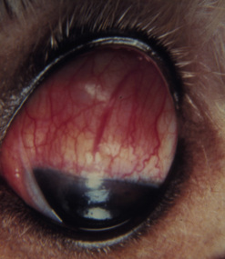
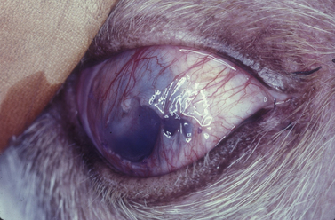
Surgical treatment for symblepharon


Symblepharon is the adhesion of bulbar to palpebral conjunctiva, the adhesion of the palpebral conjunctiva to the cornea, or the adhesion of the bulbar conjunctiva to the cornea. Symblepharon can also involve the nictitating membrane. Symblepharon is rare in dogs, but more frequent in cats. In horses, symblepharon may follow trauma, and either no surgical correction or improper surgical alignment.


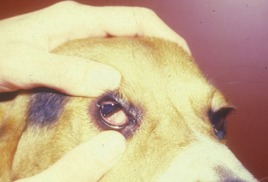
In dogs, symblepharon may develop after trauma, surgery, and chemical burns to the cornea and conjunctiva. In cats with ocular herpes (FHV-1), symblepharon may develop usually involving the cornea and the bulbar conjunctiva, the palpebral conjunctiva, or a combination of both conjunctivae (Fig. 7.7). Symblepharon associated with FHV-1 can be corrected successfully in cats, but since these corneal and conjunctival inflammations are often chronic, symblepharon formation may recur. Symblepharon affecting the cornea produces disfigurement and, if extensive, impairment of vision. Adhesions involving the bulbar and palpebral conjunctivae may shallow the conjunctival fornix, impair the drainage of tears, produce chronic conjunctivitis, and retard ocular motility.


The objectives for the surgical correction of symblepharon are to excise the fibrous adhesions between the conjunctiva and cornea, and to restore viable epithelial surfaces to the palpebral and bulbar conjunctivae, and to the cornea. Various conformers, symblepharon lenses, and silicone strips are available to physically separate the healing conjunctival surfaces and help establish and maintain the conjunctival fornix. These temporary implants are generally retained in position by complete temporary tarsorrhaphies for a few weeks for the epithelial healing to be completed. If any of these corneal or conjunctival structures is not covered with epithelium postoperatively, adhesions will recur.


After general anesthesia, clipping of the eyelid hair, and surgical preparation of the eyelids, the area is draped for aseptic surgery. The conjunctiva is thoroughly cleaned with sterile saline and all foreign material removed by cotton-tipped applicators. After placement of a wire speculum to retract the eyelids, the conjunctiva adhered to the cornea is removed by superficial lamellar keratectomy. The periphery of the corneal lesion is incised by the Beaver No. 6400 microsurgical blade to the level of the superficial stroma (Fig. 7.8a). After lifting the edge of the incision with thumb forceps with 1 × 2 fine teeth jaws, the adherent conjunctiva is excised from the corneal surface (Fig. 7.8b). If the symblepharon continues into the conjunctiva, the incision is continued and the affected conjunctiva excised (Fig. 7.8c). Once the conjunctiva is freely moveable, its edge is apposed to the limbus with 5-0 to 7-0 simple interrupted absorbable sutures. If a defect remains in the bulbar and/or palpebral conjunctiva, its edges are apposed with 5-0 to 7-0 simple interrupted absorbable sutures. To cover the healing cornea and prevent the development of new adhesions between the cornea and conjunctiva, a plastic methyl methacrylate corneal protector (Crouch corneal protector; Storz, St Louis, MO) may be inserted or amniotic membrane apposed by sutures. A soft corneal contact lens may be used instead of the thicker corneal protector (Fig. 7.8d). If considerable adhesions are present between the bulbar and palpebral conjunctivae, a thin strip of silicone sheeting is fashioned to fill the area and secured in position with 4-0 to 7-0 simple interrupted non-absorbable sutures as well as 5-0 to 7-0 simple mattress sutures placed through the silicone strip and the full-thickness eyelid with the suture knots on the external lid surface (Fig. 7.8e). To retain the corneal contact lens and reduce eyelid movements, a partial temporary tarsorrhaphy is performed with 4-0 to 6-0 simple mattress sutures positioned at one-half thickness of the eyelids (Fig. 7.8f). For details on how to perform the temporary tarsorrhaphy, see Chapter 5. After recovery from general anesthesia, an E-collar is placed on the animal to prevent self-mutilation of the surgical site.


Postoperatively, topical antibiotic solution is instilled on the eye four to six times daily. Systemic antibiotics are also administered. Once corneal epithelialization is complete, as evidenced by the lack of topical fluorescein, topical corticosteroids are added to reduce scar tissue formation. After 2–4 weeks, the tarsorrhaphy sutures are released to remove the corneal contact lens. About 2 weeks later, the fornix silicone strip and sutures are removed. Topical antibiotics/corticosteroids are continued for another 7–10 days.


This procedure provides good results in dogs and cats in which the symblepharon was secondary to trauma or chemical burns, provided the contact lens is retained in position for several weeks to permit complete epithelialization of the apposing conjunctival surfaces. This method is less successful in cats when the symblepharon appears secondary to chronic feline herpes virus infections, because with recurrent or chronic FHV-1 the corneal and conjunctival epithelia may be damaged again and re-adhesion occurs.


Conjunctival grafts/transplantation


Conjunctival grafts or flaps were first performed in humans in 1860 (Teale) and 1884 (Bock) for the treatment of symblepharon. Conjunctival autografts for the treatment of corneal ulcerations in humans for the past several decades have been replaced by partial- and full-thickness keratoplasty. The major reason is the high visual acuity in humans and the absolute need for a clear cornea. Nevertheless, conjunctival autografts are still used in humans for fungal keratitis, selected herpes simplex ulcerations, and chemical burns of the cornea.


Reports of bulbar and palpebral conjunctival grafts and conjunctival keratoplasty first appeared in the veterinary medical literature more than 50 years ago (Uberreiter 1937, Shuttleworth 1939, Stern 1950, Livingston 1950, Henderson 1951, Dimic 1957, and Berge and Westhues 1956). Most reports described advancement bulbar conjunctival grafts, complete bulbar conjunctival grafts, and palpebral conjunctival grafts. In veterinary ophthalmology the routine use of keratoplasty has not yet occurred, but the development of conjunctival autografts for the surgical management of corneal ulceration has been continuously refined. As a result, more transparent corneas now result. Further improvements may follow in the future using porcine small intestinal submucosa (available commercially) and amniotic membranes (not available commercially), rather than conjunctival grafts.


Because the veterinary ophthalmologist is concerned first for the preservation of the globe and second for clinical vision in animals, the cornea need not be perfectly and totally clear. As each animal has ample bulbar and palpebral conjunctivae for temporary or even permanent transplantation to the cornea, availability and host acceptance are not limiting factors. Treatment of deep corneal ulcerations, descemetoceles, and perforated corneal ulcers with conjunctival autografts in small animals usually halts progression and initiates healing of the corneal ulcer, transplants epithelium, fibroblasts and blood vessels to a weakened cornea, and maintains vision.


Conjunctival autografts


Conjunctiva to conjunctiva


Conjunctival autografts are used infrequently, because canine and feline conjunctival defects usually heal by secondary intention, and transposition to other areas in the same eye or between eyes is seldom indicated. Nevertheless, destruction of large areas of the conjunctiva after trauma, chemical burns, and loss after conjunctival neoplasm excision may require transposition of conjunctival tissues from other conjunctival sites.


Thin mucosal grafts can be easily constructed from the dorsal bulbar conjunctiva because this is the most accessible and the largest source. Most grafts are performed free-hand, because the conjunctival defects are usually irregular in shape and size. Transplantation to the ventral bulbar and palpebral conjunctival areas is more difficult, and construction of the ventral fornix requires long-term conformers. Grafts from the dorsal bulbar and palpebral conjunctivae are more convenient. The upper conjunctival fornix provides primarily for ocular mobility, but not for the collection and maintenance of tears.


Conjunctival autografts are performed under general anesthesia and routine surgical preparation of the eyelids and conjunctival surfaces. Conjunctival grafts must be thin and devoid of most of the underlying connective tissues. Most conjunctival grafts are either free-hand island or pedicle types. Pedicle grafts are preferred if sufficient adjacent conjunctiva is available. Mucous membrane grafts should be free of pigmentation. The conjunctival graft site must be carefully prepared, and any necrotic or potentially infected tissues removed. The adjacent bulbar conjunctiva is incised by small tenotomy scissors to produce a pedicle flap to cover the surgical defect (Fig. 7.9a). The thin conjunctival pedicle should be 1–2 mm larger than the graft site to compensate for graft shrinkage. As the scissors undermine and separate the conjunctival mucosa from Tenon’s capsule, the scissors’ tips should be plainly visible when the graft is sufficiently thin. Once fitted to the graft site, the edges of the graft and conjunctival mucosa are carefully apposed to ensure epithelium to epithelium apposition with 5-0 to 7-0 simple interrupted absorbable sutures (Fig. 7.9b). A partial temporary tarsorrhaphy can be used to decrease eyelid trauma, and provide pressure to facilitate apposition of the graft to the underlying Tenon’s capsule.


Conjunctival autografts to cornea


Conjunctival autografts are frequently used in small animal ophthalmology in clinical management of deep corneal ulcers, descemetoceles, and perforated corneal ulcers (Figs 7.107.12). Conjunctival autografts consist of either bulbar or palpebral conjunctival mucosa with epithelium and connective tissue (fibroblasts, blood vessels, and lymphatics). These autografts can be transposed and sutured onto the cornea to provide additional support and tissue for a cornea weakened by deep ulceration, descemetocele, or perforation with or without iris prolapse. The transplanted conjunctival autograft provides additional tissues and no risk of host rejection.
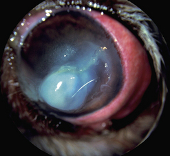


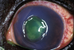
Conjunctival autografts provide sufficient tissue to strengthen a weakened cornea and prevent staphyloma formation. If additional strength is indicated, a frozen section of sterile cornea or sclera is positioned in the corneal defect before the conjunctival graft is applied. Conjunctival grafts provide new and often highly viable epithelium. When harvested from the limbal area, the transplanted conjunctival epithelium is also a stem cell capable of additional generation and transition into corneal epithelium. The conjunctival autograft contains blood vessels and lymphatics to offer significant antibacterial, antifungal, antiviral, antiprotease, and anticollagenase effects. With conjunctival transplants, leukocytes, antibodies, serum, and α2-macroglobulin (thought to be the anticollagenase factor) are immediately incorporated into the corneal ulcer bed. Because of the conjunctival blood vessels, systemic antibiotics can enter the ulcer site in higher levels. The fibrovascular or deeper layer of the conjunctival transplant offers immediate fibroblasts and collagen to begin rebuilding the corneal stroma (Fig. 7.13).
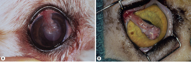


Conjunctival autografts from either bulbar or palpebral conjunctiva should be thin, and not include Tenon’s capsule or the bulbar fascia. The inclusion of Tenon’s capsule creates a thicker than necessary graft, and may contribute to surgical failure by increasing tissue contraction and tension by the transplanted conjunctiva. The scar will be more prominent with a thick autograft. Transpalpebral conjunctival autografts contain limited portions of the fibrous tarsal layer which may be necessary to maintain the graft base from the deeper aspects of the eyelid to the corneal surface. As a general guide, if the surgeon can visualize the ophthalmic scissors beneath the conjunctival graft as it is being prepared, the graft is sufficiently thin.


Conjunctival autografts are more difficult to perform than nictitating membrane flaps, but are easier than corneoconjunctival and corneoscleral transpositions, and the different types of keratoplasty procedures. Conjunctival autografts are indicated for progressive and medically non-responsive corneal ulcers, fungal corneal ulcers, deep stromal corneal ulcers, descemetoceles, ‘leaking’ corneal ulcers (positive Seidel test), perforated corneal ulcers, and perforated corneal ulcers with iris prolapse.


Some ophthalmic instrumentation is essential to perform these grafts and usually includes: an eyelid speculum, Beaver No. 6400 or 6700 microsurgical blade, Beaver scalpel handle, small tenotomy or Steven’s scissors, both small serrated and 1 × 2 teeth thumb forceps to handle the conjunctiva, ophthalmic needle holder usually with a lock (to accommodate 5-0 to 7-0 ophthalmic sutures), and suture tying thumb forceps. Some magnification (5–10×) for the operative procedure is highly recommended.


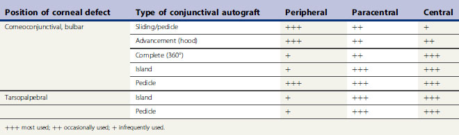
There are several different types of conjunctival autograft (Table 7.1). The divisions are based on the source of the mucosa (bulbar, tarsopalpebral, or corneoconjunctival) and the type of graft (advancement, bridge, complete, island (free), or pedicle). The dorsal bulbar conjunctiva is the most frequent source of mucosa, because of its accessibility and large surface area. The transpalpebral graft is usually constructed from the upper eyelid and sufficient tissue is available for any size corneal defect. Generally, the more central the corneal defect, the more critical the conjunctival grafting procedure.


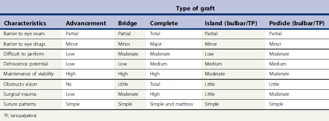
The different types of conjunctival autograft have different clinical characteristics that influence their clinical use (Table 7.2). The larger the surface area of the cornea covered by the conjunctival graft, the greater the postoperative impairment to patient’s vision, the greater the barrier to postoperative intraocular examination, and, at least theoretically, the greater impediment for the corneal and intraocular penetration of most ophthalmic drugs. In those types of graft used to cover the central cornea, and for the more serious corneal ulcers, these techniques are often more difficult to perform. Attachment of the conjunctival graft to the progressive central corneal ulceration must be exact. Magnification provided by a head loupe or preferably the operating microscope is necessary for those types of conjunctival autograft that are apposed by sutures directly to the adjacent corneal epithelium and stroma.


Preparation for surgery for the different types of conjunctival autograft is similar. Once under general anesthesia, the eyelids are carefully clipped and the eyelid skin prepared for surgery. The conjunctival surfaces are carefully cleaned of any debris with sterile cotton-tipped applicators. Dilute solutions of 0.5% povidone–iodine are used to treat the surfaces of both the conjunctiva and cornea to reduce the overall microbial population, and then rinsed from the eye with sterile saline. A lateral canthotomy may be indicated to increase exposure of the surgical site and facilitate the surgery for most dogs except the brachycephalic breeds.


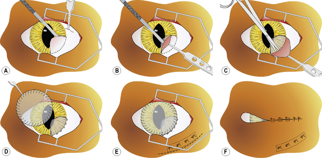
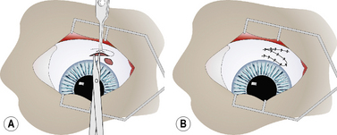
Complete (360°) bulbar conjunctival autograft (Gundersen type)


The complete, 360°, or Gundersen-type conjunctival autograft has been used extensively in veterinary ophthalmology since its first description nearly 50 years ago, but has now been partly replaced by conjunctival grafts that only partially cover the cornea. In this graft nearly all of the bulbar conjunctiva is separated from the underlying Tenon’s capsule to cover the entire cornea. The graft covers the corneal defect but is not apposed directly by sutures. Because the entire cornea is covered, patient vision, examination of the eye, and the intraocular penetration of topical drugs through both the vascularized mucosa and the cornea is reduced. With this graft, systemic as well as topical administration of drugs is recommended. Of all of the different types of conjunctival graft, the complete 360° conjunctival graft provides the maximum support for the entire cornea. Corneal defects involving the central and paracentral areas of the cornea are treated with this type of conjunctival graft.


For the 360° fornix-based conjunctival graft, the dorsal bulbar conjunctiva is elevated by fine teeth thumb forceps and incised by scissors at the limbus (Fig. 7.14a). The bulbar conjunctiva is separated from the underlying Tenon’s capsule by alternating blunt–sharp dissection by small tenotomy scissors with blunt tips. For a reasonably thin conjunctival graft, the scissors’ tips should be easily observed through the thin mucosa (Fig. 7.14b). To facilitate dissection, saline can be injected subconjunctivally to help separate the bulbar conjunctiva from Tenon’s capsule. Some hemorrhage is expected and depends on the extent of conjunctival hyperemia associated with the corneal ulceration and secondary iridocyclitis. If the surgical dissection plane enters Tenon’s capsule, additional hemorrhage results.
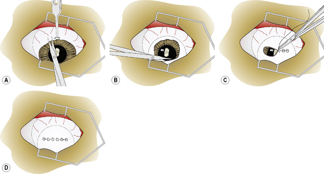


The bulbar conjunctiva is dissected for 360° about the limbus. The most difficult area is usually under the nictitating membrane (Fig. 7.14b). As the cornea measures about 15 × 16 mm in the dog, and 16 × 17 mm in the cat, in vertical and horizontal diameters, respectively, adequate amounts of bulbar conjunctiva necessitate 8–10 mm of dissection from the limbus for 360°. As the different rectus muscles in the dog and cat insert 6–10 mm from the limbus, preparation of the conjunctival graft requires surgical dissection immediately beneath the bulbar conjunctiva and not on the sclera.The conjunctival graft must be thin to minimize traction and excessive pressure on the sutures postoperatively. When properly prepared, the loosened edges of the bulbar conjunctival graft should rest on the central cornea and not retract spontaneously to the limbus.


The edges of the bulbar conjunctiva are apposed horizontally with 5-0 to 7-0 absorbable simple interrupted or simple mattress sutures (Fig. 7.14c). Usually four to six sutures are necessary to appose the dorsal and ventral conjunctival edges. Simple interrupted mattress sutures are recommended if the graft is thicker than desirable or additional traction on the suture line is anticipated. A purse-string stitch has also be used but is not recommended as this produces additional tension on the graft as all edges are pulled to the center of the cornea. Once completed, the 360° bulbar conjunctival graft covers the entire cornea (Fig. 7.14d).


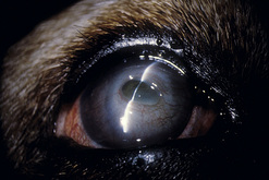
The transposed conjunctival mucosa is not usually sutured directly to the corneal defect, but can be if perforation is likely or the deep corneal ulcer is already leaking aqueous humor (Fig. 7.15). As these grafts completely cover the cornea, vision in the eye is obscured and intraocular inspection is not possible.


Stay updated, free articles. Join our Telegram channel

Oct 14, 2016 | Posted by in SUGERY, ORTHOPEDICS & ANESTHESIA | Comments Off on Surgical procedures for the conjunctiva and the nictitating membrane

Full access? Get Clinical Tree

Get Clinical Tree app for offline access