Surgery of the eyelids

Chapter 5 Surgery of the eyelids


Introduction


Eyelid diseases are common in dogs and horses, and infrequent in cats and cattle. In contrast to most ophthalmic diseases, the initial clinical management of eyelid disorders is usually surgery. Traditionally, eyelid diseases are divided into congenital and developmental, inflammatory, traumatic, and neoplastic. The clinical management of all of these groups of eyelid diseases, except for the inflammatory types, is surgery. Surgical treatment may also be indicated for the inflammatory eyelid diseases, after resolution with antimicrobial therapy, to restore the eyelid contours and function associated with the excessive postinflammatory scarring and distortion.


Anatomy


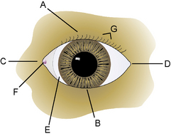
The morphology of the eyelids in domestic animals is quite similar, with size being the major variable. The eyelids represent the transition of the integument system and the beginning of the ophthalmic apparatus with the initiation of the palpebral conjunctiva. The eyelids surround the palpebral fissure, through which the eye contacts the environment. The eyelids are divided clinically into the dorsal, superior, or upper eyelid; the ventral, inferior, or lower eyelid; the medial or nasal canthus; and the lateral or temporal canthus (Fig. 5.1). The dorsal eyelid is the largest, most mobile, and 2–5 mm longer than the lower lid. Distinct ligaments, the septum orbitale, and certain muscles attach at both sides of the palpebral fissure, resulting in an oval rather than round eyelid opening. The medial canthal eyelid area is relatively fixed to the subcutaneous tissues and periosteum by the medial palpebral or canthal ligament. Protected immediately behind the medial canthal ligament is the nasolacrimal sac. The lateral canthal region is more mobile, especially in dogs. The lateral canthal ligament is poorly developed in the dog and is replaced by the retractor anguli oculi lateralis muscle. Hence in dogs, defects often affect the lower lid and lateral canthus.


Eyelid skin, cilia (eyelashes), and glands


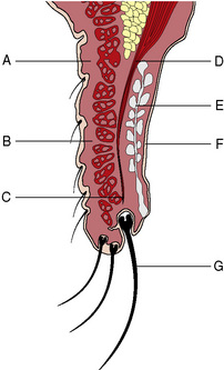
The skin of domestic animal eyelids is thinner than other parts of the integument system (Fig. 5.2). Eyelid movements require both thin and pliable skin. Fine short hairs normally cover the eyelid skin. The subcutaneous tissues under the eyelid skin are relatively thin and attach the lid skin to the deeper orbicularis oculi muscle. Cilia or eyelashes occur primarily on the canine upper eyelid, usually in two or four irregular rows. These cilia are usually the same color as the adjacent eyelid hair coat. Long tactile hairs (pili supraorbitales or vibrissae) appear as a tuft along the dorsal medial orbital margin in several animal species. Eyelashes are not present in cats, although the eyelid hair next to the dorsal eyelid margin may be considered a substitute. Two types of gland, the glands of Moll and glands of Zeis, are located about the cilia follicles. The gland of Moll is a modified sweat (apocrine) gland and the gland of Zeis is a modified sebaceous gland. These glands can become inflamed and abscessed in young animals, resulting in the formation of a stye or external hordeolum. The margo-intermarginalis represents the free margin of the eyelids.


Eyelid muscles


The second layer of the eyelids is the muscle layer which consists of several muscles that either close or open the palpebral fissure. The orbicularis oculi muscle is the predominant muscle involved in closure of the palpebral fissure in domestic animals. This muscle encircles the entire palpebral fissure and is attached by the septum orbitale to the medial and lateral canthi. It is divided into the inner pars palpebralis and the outer pars orbitalis. Both the origin and insertion of the orbicularis oculi muscle is the medial palpebral ligament. This muscle is immediately beneath the skin layer, and surgical procedures for entropion (inversion of the eyelid margin) and ectropion (eversion of the eyelid margin) directly involve this muscle. In large animal species the orbicularis oculi muscle is very powerful, especially when ophthalmic pain is present. In large animals, local nerve blocks of the palpebral nerve branch of the auriculopalpebral nerve (branch of the facial nerve) is often necessary to relax this muscle to adequately examine and even treat a painful eye.


Several muscles are involved in opening the eyelids and increasing the size of the palpebral fissure in the dog and cat. In the canine upper eyelid these muscles consist of (medial to lateral) the levator anguli oculi medialis, levator palpebrae superioris, and the frontalis, and in the lower lid, the pars palpebralis of the sphincter colli profundus (Fig. 5.3). The levator palpebrae superioris muscle has its origin deep within the orbit, along with the rest of the extraocular muscles, and lies immediately above the dorsal rectus muscle that inserts several millimeters from the dorsal limbus of the globe. Fascial attachments between the dorsal rectus and levator palpebrae superioris muscles result in simultaneous upper movements of the globe and retraction of the upper eyelid. The levator palpebrae superioris muscle seems to be the most important muscle for upper lid retraction as damage to this muscle or its insertions into the tarsal layer results in ptosis (or drooping of the upper eyelid). The levator anguli oculi medialis muscle elevates the medial upper eyelid and erects the long tactile hairs of the eyebrow. In some of the large breeds of dogs, the action of this muscle results in a noticeable notch at the junction of the medial and middle one-thirds of the upper lid. In the dog, Müller’s smooth muscle fibers, innervated by the sympathetic nerves, attach to the upper tarsus; in the cat, these muscle fibers also attach to the nictitating membrane. With the release of endogenous adrenaline (epinephrine), or during the fight-and-flight reflex, these adrenergic innervated smooth muscle fibers can immediately increase the size of the palpebral fissure.


In the cat, the major muscle for closing the palpebral fissure is the orbicularis oculi. The corrugator supercilii medialis raises the majority of the upper eyelid, but laterally the frontoauricularis helps to elevate the upper lid. Lateral and as a substitute for the lateral canthal ligament, the corrugator supercilii lateralis elongates the palpebral fissure. The lower eyelid is relatively fixed and only the orbicularis oculi is present to close the palpebral fissure.


In the horse, the anatomy of the eyelids follows the other mammalian patterns. The upper lid is considerably larger than the lower lid and contributes most of the lid motility. The lacrimal puncta are about 2 mm in diameter and about 8 mm from the medial canthus. The medial canthus can be prolonged medioventrally and possesses a prominent, occasionally pigmented, caruncle. The upper lid possesses about 40–50 meibomian glands dorsally and 30–35 glands ventrally.


In cattle, the eyelids are quite similar to the horse. They are very thick and strong! The orbicularis oculi is often in small bundles and extends to the edge of the lid margin. The orbicularis oculi is attached medially to the strong medial palpebral ligament and lacrimal bone. Although cattle possess a small and weak lateral palpebral ligament, entropion and ectropion occur rarely in cattle. There are about 32–34 meibomian glands in the upper lid and 26–28 in the ventral lid. The upper fornix measures about 36 mm from the upper lid margin, and the lower fornix is 22 mm from the lower lid margin.


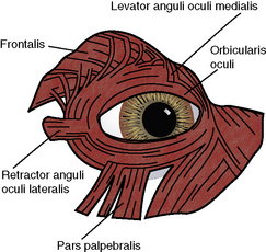
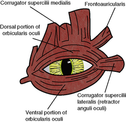
The lateral palpebral or canthal ligament is poorly developed in dogs and cats, and usually consists of an irregular thickened lateral septum orbitale. In the large breeds of dogs the lack of or a poorly developed lateral canthal ligament contributes directly to lateral canthal lid diseases in these breeds. The major component of the lateral canthal support system has been replaced by the retractor anguli oculi lateralis (dog) or corrugator supercilii lateralis (cat) muscle (Fig. 5.4). This results in a somewhat mobile but unstable lateral canthus in dogs, and contributes to the frequent involvement of the lateral canthus and lateral lower and upper eyelids with entropion and ectropion. The lateral one-half of the lower eyelid in the dog also has the pars palpebralis muscle, a subdivision of the sphincter colli profundus muscle, which can depress the lateral lower lid.


Eyelid tarsal layer and glands


The eyelid skin and tightly adherent muscle layers are easily separated surgically from the deeper two layers, the fibrous tarsus containing the tarsal or meibomian glands and the inner palpebral conjunctiva. The fibrous tarsus provides some infrastructure for the eyelids, but not to the extent that the tarsal hyaline plate does in humans. The fibrous tarsus has fascial attachments to the septum orbitale, resulting in a strong connection to the periosteum of the orbital rim, and a significant barrier for trauma, surgery, and external infectious agents to enter the orbit. The fibrous tarsal layer is also in intimate contact with the medial palpebral or canthal ligament, the base of the nictitating membrane, and the lateral canthus. The medial palpebral ligament is more distinct than the lateral ligament in the dog, and consists of a fibrous band originating from the periosteum of the frontal bone that inserts into the upper and lower tarsal layers. The medial palpebral ligament also serves as the origin and insertion for the circular orbicularis oculi muscle, which undoubtedly assists in the medial movement of tears on the cornea and within the conjunctival sacs. In the middle section of the upper eyelid the levator palpebrae superioris muscle inserts into the tarsal layer. The superior and inferior tarsal muscles are smooth muscles in the dog within the endorbita that inserts into the tarsus. These muscles are under sympathetic innervation and help maintain the palpebral fissure open.


The tarsal or meibomian glands are sebaceous (holocrine) types and produce the important outer lipid or oily fraction of the preocular or precorneal film. The lipid layer of the preocular film prevents evaporation of the thicker aqueous layer and stabilizes the preocular film. The number of tarsal glands in each lid ranges from 20 to 40, with the upper eyelid containing more glands. The orifices of the tarsal or meibomian glands empty onto the center of the eyelid margin. This area is referred to the ‘gray line’ and is an important surgical zone. The tarsal glands are occasionally visible through the palpebral conjunctiva, and extend for 3–5 mm into the lid substance. The tarsal glands seem able to undergo metaplasia and form additional cilia, called distichia. At the medial canthus at the junction of the upper and lower lids, and at the base of the nictitating membrane, is the lacrimal caruncle. Small fine hairs emerge from its surface, which can act as a wick for tears to moisten the medial canthal skin. The lacrimal caruncle also contains small sebaceous glands.


Anatomy of the equine eyelid


Muscles that open the equine eyelids include the levator palpebrae superioris, Müller’s, levator anguli oculi medialis, frontalis, and malaris. The levator palpebrae superioris muscle, along with the levator anguli oculi medialis muscle, raises the upper eyelid. Müller’s muscle is a smooth muscle that originates from the posterior surface of the levator muscle in the upper eyelid and from the ventral rectus muscle for the lower eyelid. It is innervated by sympathetic fibers that travel with the ophthalmic branch of the fifth cranial nerve. Müller’s muscle inserts on the tarsus and, along with other muscles, keeps the palpebral fissure open. It is the muscle, when innervation is interrupted in Horner’s syndrome, that results in ptosis of the upper eyelid. The frontalis muscle inserts laterally on the upper eyelid. The malaris muscle inserts on the ventral orbicularis oculi muscle and functions to open the lower eyelid. The upper eyelid is the more mobile and larger section of the eyelids. It provides the majority of the blinking function.


Sensory innervation of the eyelids is via the ophthalmic and maxillary branches of the trigeminal (fifth cranial) nerve. The ophthalmic portion branches into the frontal, lacrimal, and nasociliary nerves. The frontal branch passes anteriorly from the orbit through the supraorbital foramen. It then becomes the supraorbital nerve, dividing over the forehead and innervating most of the upper eyelid. The lacrimal nerve innervates the lacrimal gland and the upper eyelid at the lateral canthus. The nasociliary branch gives rise to the infratrochlear nerve, which provides sensory innervation to the medial canthus, caruncle, nictitating membrane, upper and lower conjunctiva, and nasolacrimal puncta and ducts. The remainder of the lower eyelid is innervated by the zygomatic nerve, which is a branch of the maxillary nerve.


Preoperative examination procedures


In the assessment of the eyelids preoperatively, their structure, function (blink reflex), and relationship to the face, each other, and to both eyes are carefully evaluated. Adequate illumination and some magnification are essential. The head loupe or magnifier and Finoff transilluminator, or portable slit-lamp biomicroscope are the best instruments that combine these two characteristics. The eyelids, eye, and orbit relationships can also be influenced by the presence of pain, inflammation, enophthalmia or exophthalmia, body condition, age, dehydration, and muscle condition. Not infrequently, the eyelids cannot be restored surgically to completely normal appearance and function because of these other variables, especially the position of the globe. These complex relationships of the eyelids, eye, and orbit in the dog also impede genetic studies of the eyelid diseases. The animal eyelids normally rest on the cornea and bulbar conjunctiva. If the globe is recessed into the orbit, eyelid contact may not be possible and instability of the lower eyelid results. The usual result is entropion or an inversion of the eyelid margin and substance. This phenomenon occurs commonly in certain breeds of dogs, foals, and in aging animals (probably associated with loss of orbital fat).


Secondary blepharospasm is a common component of most painful eyelid conditions. With eyelid swelling, the eyelid margin usually rolls inward. When the outer eyelid margin, normal cilia (eyelashes), and trichiasis (involving the normal eyelid hair) touch the bulbar conjunctiva, cornea or a combination of both surfaces, the animal reacts by developing secondary blepharospasm. The resultant trigeminal–facial nerve reflex usually worsens the eyelid defect even further, and produces additional irritation and pain. This protective eyelid reflex then produces an ever-increasing cycle of pain and blepharospasm. Hence, in many painful eyelid diseases, the initial structural disease is aggravated by a normally protective eyelid closure reflex. Animals often respond to localized ocular pain by rubbing, which can cause additional localized swelling and even loss of skin integrity. Therefore, in the examination of eyelid diseases that are potential candidates for surgical correction, surgery should be directed at only the underlying structural eyelid disorder.


To estimate the extent of secondary blepharospasm in a patient, a few drops of topical anesthetic are instilled onto the cornea and conjunctiva after the initial entropion has been estimated. After 3–5 min, the secondary blepharospasm will usually be relieved, and the basic structural eyelid abnormality can be ascertained. Surgical correction should be directed at only this anatomic eyelid abnormality. The defect is usually undercorrected slightly (about 0.5–1 mm) to accommodate postoperative fibrosis.


Infrequently, the eyelid defect can become so painful and the eyelid and associated tissues so inflamed that multiple instillations of topical anesthetic will not totally suppress secondary blepharospasm. In these patients, localized regional eyelid block of the palpebral nerve can be administered.


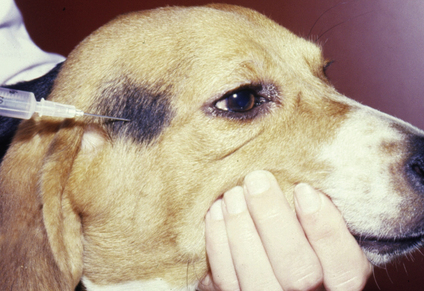
In the dog, a few milliliters of local anesthetic are injected subcutaneously along the dorsal aspects of the middle portion of the zygomatic arch to block the palpebral branch of the facial nerve and the primary innervation to the orbicularis oculi muscle that closes the palpebral fissure (Fig. 5.5). Within a few minutes, total loss of eyelid muscle tone will occur, and the extent of the eyelid problem to be corrected surgically can be determined.


In the horse, the auriculopalpebral nerve can be blocked at at least two sites (see Fig. 3.5, p. 44). The auriculopalpebral nerve can be blocked by infiltrating local anesthetic in a fan-like manner subfascially in the depression just caudal to the posterior ramus of the mandible at the ventral edge of the temporal portion of the zygomatic arch. The hypodermic needle is directed dorsally just caudal to the highest point of the arch. Before injecting local anesthetic, aspiration is performed to prevent injection into the rostral auricular artery or vein. This procedure may also result in akinesia of the ear muscles as well as gravitate ventrally and affect other branches of the facial nerve. Within a few minutes, total loss of eyelid muscle tone will occur, but lid sensation is still present.


The second palpebral nerve block in the horse blocks multiple branches of the palpebral nerve closer to the eye. Local anesthetic is injected subcutaneously at the highest point of the dorsal border of the zygomatic arch.


The supraorbital nerve of the horse is often blocked at the supraorbital foramen to provide akinesia to the forward branches of the palpebral nerve, as well as local anesthesia by blocking the supraorbital nerve, a branch of the trigeminal nerve. This procedure is often used prior to ophthalmic examinations, insertion of dorsal subpalpebral medication systems, and excision of upper eyelid masses.


In cattle, the palpebral nerve can be blocked by injecting 3–5 mL of local anesthetic about 4–6 cm caudal of the lateral canthus.


Surgical considerations of the eyelids


The upper and lower eyelids share most functions, but also have some unique characteristics. The upper eyelid covers the majority of the cornea, and blinks at a normal rate of about 15 times per minute. Only part of these blink reflexes cover the entire cornea. The upper eyelid has the levator palpebrae superioris muscle that originates from the depths of the orbit above the dorsal rectus muscle. The upper eyelid is primarily responsible for the cosmetic appearance, while the lower eyelid margin serves to collect and prevent the preocular film and tears from overflowing onto the lower canthi and eyelid surfaces. Surgical and traumatic interruption of the palpebral nerve innervation to the eyelids results in exposure keratitis if upper, rather than lower, eyelid function is impaired.


The upper eyelids are larger and longer than the lower lids, and are the principal source for tissues for reconstructive surgical procedures of the eyelids. The higher number of tarsal or meibomian glands occurs in the upper eyelid. Only the canine upper eyelid contains cilia (eyelashes). Although the autogenous transplantation of cilia has not been reported in domestic animals, the surgical technique is not difficult. Hence, surgery of the upper eyelids must consider movements, protection of the eye, and appearance, whereas surgery of the lower eyelids must primarily address the collection, retention, and medial movement of tears. After extensive blepharoplasty, the eyelids (especially the upper) may function poorly for several days to weeks because of lid swelling and possible nerve impairment. To protect the cornea and prevent ulceration, a temporary complete tarsorrhaphy is necessary until a normal blink response returns.


The conjunctival cul-de-sac or fornix is considerably larger for the dorsal eyelid, probably to accommodate ventral rotation of the globe. The lower conjunctival fornix is more shallow but is the primary receptacle for the tears and, assisted by the intermittent movements of the orbicularis oculi muscles, gradually propels the tears medially toward the lacrimal puncta.


As indicated in an earlier section, the surgical anatomy of the eyelids is usually divided into two layers: the skin and muscle layer, and the deeper tarsus and palpebral conjunctiva. The thin elastic eyelid skin has limited subcutaneous tissue and attaches directly to the orbicularis oculi muscle. Surgical separation of the eyelid skin from the deeper muscle layer is tedious and, in small species, often very difficult. Within the eyelid margins are the numerous orifices of the tarsal or meibomian glands. This area is referred to as the ‘gray line’, an area to surgically split or divide the eyelids longitudinally, as well as for the placement of eyelid sutures that will not contact and directly damage the cornea.


Surgical instrumentation


The instrumentation for eyelid surgery usually consists of a mixture of general soft tissue instruments, as well as selected ophthalmic instruments. The recommended ophthalmic instruments include small straight and curved strabismus or tenotomy scissors to cut tissues and sutures, both teeth (1 × 2) and serrated thumb forceps (such as Bishop–Harmon forceps), small scalpel blades (Nos 6400 and 6500 microsurgical), small wire eyelid speculum, and a standard ophthalmic needle holder (often with lock). Special thumb forceps, such as chalazion and entropion forceps, are useful to clamp and stabilize the eyelids during surgery (see Table 1.3, p. 12).


Suture selection is variable and often at the surgeon’s discretion. Sutures for the tarsus and palpebral conjunctiva are usually absorbable (polyglycolic or polyglactic acid, and polydioxanone), and the simple continuous pattern is usually used. The knots should be buried beneath the palpebral conjunctiva to avoid direct contact with the cornea. Sutures involving the eyelid skin and superficial aspects of the orbicularis oculi muscle are usually non-absorbable and the simple interrupted pattern. Many veterinary ophthalmologists prefer 4-0 to 6-0 silk; if corneal contact with the silk suture occurs, no irritation or damage results. Although tissue reactivity with silk can be a problem, and braided silk is a potential wick for bacterial invasion, eyelid suture removal at 7–10 days postoperatively effectively avoids these potential problems. The other frequently used non-absorbable suture is 4-0 to 6-0 nylon. Choice of atraumatic swaged-on cutting needles is quite variable; however, the one-fourth to three-eighths curved needles are most useful.


Hemostasis is usually provided by small curved mosquito hemostats or point cautery. Vessel ligation with absorbable sutures is infrequent as these areas may develop focal postoperative fibrosis that may influence the surgical result. Point cautery is usually preferred, but used judiciously. Sterile cotton-tipped applicators and cotton surgical sponges can effectively maintain most eyelid surgical fields clear of hemorrhage. Moistening with 1:10 000 or other dilutions of adrenaline (epinephrine) may be helpful. Hemostasis is also provided when the eyelids are grasped and held with chalazion and entropion forceps.


Surgical preparations of the eyelids


Surgical preparation of the eyelids is usually performed immediately before surgery. Sometimes the patient is on therapy with topical antibiotics or antibiotics/corticosteroids for treatment of the eyelid disorder, and the same therapy is continued immediately after surgery. The high vascularity of the eyelids promotes healing and often topical corticosteroids are administered perioperatively to control the local inflammation and swelling.


A bland petroleum ointment may be placed on the corneal and conjunctival surfaces to collect debris and hair during the surgical preparation. The ointment is carefully removed by sterile cotton-tipped applicators immediately before surgery. The eyelid hair is carefully removed by small hair clippers or shaved. The eyelid skin is thin and, if traumatized during hair removal, swelling may result.


The surgical preparation of choice is 0.5% povidone–iodine solution as contact with the cornea is not irritating. At least three scrubs are recommended to clean and reduce the local microbial population. After these scrubs, the area is liberally rinsed with 0.9% sterile saline. Alcohol and other traditional surgical preoperative measures are not recommended as contact with the cornea and conjunctiva can be very irritating and damaging to their epithelia. Draping is performed with four small cotton towels positioned around the palpebral fissure and covered with a surgical drape. Small towel clamps or bulldog clamps may be used to secure the drapes, but should be used sparingly.


Tarsorrhaphy in small animals


In the tarsorrhaphy procedure the eyelids are apposed either temporarily or permanently, and the lid apposition may include part or all of the upper and lower eyelids. In the permanent tarsorrhaphy procedure part of the eyelid margins of the upper and lower eyelids are excised and, after apposition by sutures, the eyelids should remain sealed. Complete permanent tarsorrhaphies are part of the enucleation and exenteration procedures after removal of the eye and the orbital contents. Partial permanent tarsorrhaphies are indicated to treat long-term ocular disorders, such as neuroparalytic keratitis, neurotropic keratitis, lagophthalmia, and chronic exposure keratitis.


In the temporary tarsorrhaphy technique the eyelid margins are apposed by sutures for several days to a few weeks to cover the healing cornea and/or conjunctiva. In the complete temporary tarsorrhaphy method the entire palpebral fissure is closed. In the partial technique, only part (medial, central, or lateral) of the palpebral fissure is closed, thereby permitting vision by the patient, daily inspection by the veterinarian, and topical medication of the eye. The complete temporary tarsorrhaphy is indicated clinically, in part, for the treatment of traumatic proptosis, after most orbitotomies, after many extensive eyelid procedures, after nictitating membrane flaps, after extensive conjunctival surgery, to treat premature opening of the eyelids, to help maintain collagen shields and contact lenses in place, and for the treatment of recurrent corneal erosions and other selected superficial corneal disorders. Complete temporary tarsorrhaphies are also indicated when upper eyelid function is impaired and the development of exposure keratitis is anticipated. The partial temporary tarsorrhaphy is used frequently after conjunctival and corneal surgery to reduce eyelid trauma to the surgical site, and to provide some contact and pressure to fresh grafts. Suture ends in temporary tarsorrhaphies may be left long to facilitate occasional adjustment of the suture pressure, as well as occasional loosening to open the tarsorrhaphy and inspect the eye.


The support of a weakened cornea by complete temporary tarsorrhaphy may vary by breed but it does not appear to give as much support as that provided by the nictitating membrane flap or a complete conjunctival graft. However, the complete temporary tarsorrhaphy procedure can supplement these methods to provide additional corneal support. Complete temporary tarsorrhaphy constitutes a barrier to topical medication of an eye, but the subpalpebral system can be inserted in the dorsolateral or lateral conjunctival fornix at the conclusion of surgery to ensure delivery of ophthalmic solutions. The apposed eyelids may also retain the topical solutions in contact with the cornea for longer periods of time, thereby increasing their effectiveness.


The permanent apposition of the upper and lower eyelid margins at the medial and lateral canthi is discussed in a later section under surgical procedures to reduce the size of the palpebral fissure. Chronic exposure of the cornea, especially in brachycephalic breeds, may be a major contributing factor in the pathogenesis of recurrent central corneal ulceration, and surgical reduction of the size of the palpebral fissure may significantly reduce the possibility of recurrence.


Permanent tarsorrhaphy


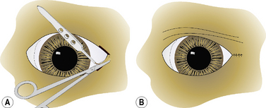
In the permanent tarsorrhaphy procedure the upper and lower eyelid margins are trimmed by curved Metzenbaum scissors 3–5 mm from the lid margins (Fig. 5.6a). This area is usually where the eyelid pigmentation stops and the eyelid hair appears. For complete permanent tarsorrhaphy the entire eyelid margins are excised for 360°, with special care to adequately excise the lid margins at the two canthi. For partial permanent tarsorrhaphy any section of the eyelid may be used, but most often the lateral and medial canthal areas are involved. At a depth of 4–5 mm, the bases of the meibomian glands are excised. The eyelids are usually apposed by two layers of sutures in most dogs (Fig. 5.6b); however, in miniature breeds, a single suture layer may suffice. The deeper tarsopalpebral layer is apposed with either 4-0 to 6-0 simple interrupted or a simple continuous absorbable suture placed submucosally to avoid corneal contact. The eyelid skin–orbicularis oculi layer is usually apposed with 4-0 to 6-0 simple interrupted or interrupted mattress non-absorbable sutures.


Stay updated, free articles. Join our Telegram channel

Oct 14, 2016 | Posted by in SUGERY, ORTHOPEDICS & ANESTHESIA | Comments Off on Surgery of the eyelids

Full access? Get Clinical Tree

Get Clinical Tree app for offline access