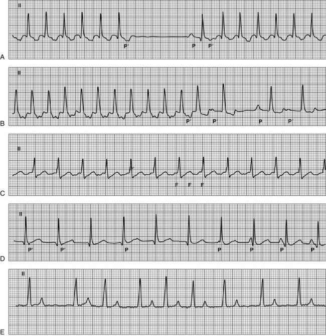
Supraventricular Tachyarrhythmias in Dogs

Chapter 171


Supraventricular Tachyarrhythmias in Dogs


Definition


Tachyarrhythmias are rapid rhythms, with rates typically above 180 to 200 beats/min in dogs, that appear out of proportion to the animal’s level of activity or stress. This chapter outlines the diagnostic approach and management strategies for common and uncommon supraventricular tachyarrhythmias (SVTs) in dogs. Additional information regarding management of focal atrial tachycardias, atrial flutter, and atrial fibrillation can be found in Chapters 175, 176, and 178.


Tachyarrhythmia management is based on the discrimination between supraventricular and ventricular rhythms. SVTs have in common that their maintenance necessitates the participation of at least one cardiac structure situated above the ventricles. These structures include the sinus and atrioventricular (AV) nodes, the bundle of His, the atrial myocardium, the tributary veins such as the coronary sinus, the pulmonary veins or venae cavae, and on occasion accessory pathways. The main characteristic of SVTs on the surface electrocardiogram (ECG) is a narrow QRS complex (<70 ms in dogs), which indicates that electrical impulses, once they reach the ventricles, propagate within specialized muscular bundles of the His-Purkinje conduction system, similar to normal sinus beats. However, it is not unusual for SVTs to display broader QRS complexes as a result of an anatomic lesion or a rate-dependent prolongation of intraventricular conduction (e.g., bundle branch block).


Pathophysiology


The initiation of arrhythmias requires precipitating factors and a suitable substrate. Supraventricular structures such as vascular orifices opening into the atria (cranial and caudal vena cava, coronary sinus, pulmonary veins), muscular ridges (crista terminalis, eustachian ridge), and valve annuli create areas of block, slow conduction, and tissue boundaries that facilitate arrhythmia wave front propagation. Although myocyte depolarization is usually limited to the sinus and AV nodes, other supraventricular structures, in particular the crista terminalis and pulmonary veins, can become sources of focal ectopic activity. Ischemia, interstitial fibrosis, inflammation, and atrial chamber dilation also contribute the arrhythmogenic substrate. Additionally, autonomic influences and electrolyte abnormalities can contribute to the initiation and perpetuation of arrhythmias. It is therefore understandable that heart failure is frequently accompanied by SVTs.


Electrophysiologic mechanisms responsible for SVTs can be categorized into abnormalities of electrical impulse formation and abnormalities of impulse propagation. The first group of mechanisms encompasses abnormal automaticity in which myocytes depolarize spontaneously at a faster rate than sinus node cells and triggered activity in which spontaneous depolarization of cells is precipitated by repolarization abnormalities from the preceding action potential. Another mechanism known as reentry is the repetitive propagation of an impulse around an area of nonconducting tissue, such as a vein, a valve annulus, or a fibrotic region.


Classification


SVTs, currently diagnosed by electrophysiologic studies in dogs, can be classified according to the site of origin and the underlying arrhythmogenic mechanism. They include atrioventricular reciprocating tachycardia, focal atrial tachycardia, macro-reentrant atrial tachycardia or atrial flutter, focal junctional tachycardia, and atrial fibrillation. Although atrioventricular nodal reentrant tachycardia is a common cause of paroxysmal tachycardia in people, it has not been identified in dogs.


Atrioventricular reciprocating tachycardias (AVRTs) are macro-reentrant arrhythmias in which the electrical wave front moves along an anatomic circuit formed by the AV-His-Purkinje system, the ventricles, accessory pathways, and the atria. Accessory pathways are congenital muscular bundles that do not regress during the formation of the cardiac skeleton. These pathways represent an alternative route of AV or ventriculoatrial (VA) conduction besides the AV node. In the dog, accessory pathways are predominantly right-sided in the posteroseptal and right posterior area of the tricuspid annulus (in human nomenclature), rarely are multiple, and frequently present with unidirectional retrograde conduction (70% of cases) with no evidence of ventricular preexcitation during sinus rhythm. Two forms of AVRT have been described in human patients: orthodromic, in which the wave front descends from the atria to the ventricles along the AV node and returns to the atria along the accessory pathway, and antidromic, in which the wave front circles in the opposite direction. Only orthodromic AVRT and a preexcited form of atrial fibrillation have been described thus far in dogs.


Focal atrial tachycardias (FATs) correspond to the rhythmic atrial activation of a small area of atrial tissue (focus) from which wave fronts spread out centrifugally. In dogs, FATs are more commonly right sided (63% of cases) and automatic (78% of cases). It has been observed that FATs arising from the pulmonary veins trigger atrial fibrillation in 25% of the dogs examined.


Atrial flutter (AFL) is a macro-reentrant atrial tachycardia characterized by an organized atrial rhythm with a rate typically above 300 beats/min. The anatomic circuit of AFL includes the cavotricuspid isthmus, which is an area of the low posterior right atrium delimited by the caudal vena cava and the eustachian ridge posteriorly and the tricuspid valve annulus anteriorly. Based on the rotation pattern around the tricuspid valve annulus, two forms of cavotricuspid isthmus–dependent AFL have been identified: typical and reverse typical. During typical AFL the activation wave front turns counterclockwise around the tricuspid valve annulus when viewed from above, whereas during reverse typical AFL it turns clockwise.


Focal junctional tachycardia (FJT) is caused by an abnormally rapid discharge from the junctional region just posterior to the AV node and the bundle of His.


Atrial fibrillation (AF) is a rhythm disturbance characterized by uncoordinated atrial activations with consequent deterioration of atrial mechanical function and an irregular ventricular response rate.


Other features widely used in the human literature to characterize SVTs include their clinical presentation, duration, and mode of onset and termination. Accordingly, SVTs can be described as nonsustained (<30 seconds in duration), sustained >30 seconds in duration or causing syncope), incessant (lasting >12 hours a day), repetitive (runs of nonsustained tachycardia alternating with period of normal sinus rhythm), or paroxysmal (abrupt initiation and termination). A classification specific to AF differentiates it into paroxysmal (self-limiting and lasting <7 days), persistent (uninterrupted, but sinus rhythm can be restored with electrical or pharmacologic cardioversion), or permanent (sinus rhythm cannot be restored).


As suggested by the brief preceding discussion, SVTs are in many ways more complicated to classify and diagnose than ventricular tachycardias. Except in straightforward cases, such as persistent or permanent AF, the veterinarian should consider consultation or referral to a cardiologist because advanced methods of diagnosis and therapy might be necessary.


Clinical Manifestations and Evaluation


Common signs associated with SVT include weight loss, lethargy, exercise intolerance, excessive panting, and dyspnea. However, it is not uncommon for dogs to be asymptomatic. Owners of dogs with SVT may report seeing the dog’s heart pounding in the chest during episodes of tachycardia. Episodic weakness and transient loss of consciousness are less common with SVT, unless the vasomotor reflex is inadequate. Additionally, SVTs may be recognized only at the time of physical examination in dogs with signs of congestive heart failure. In these situations, it is difficult to determine the contribution of the arrhythmia to the clinical signs and to the degree of myocardial dysfunction because an uncontrolled ventricular rate can lead to progressive myocardial failure independent of any underlying structural disease. This phenomenon is known as tachycardiomyopathy, and it is likely that a sustained rate above 250 beats/min will induce severe myocardial dysfunction within 3 to 4 weeks. It is noteworthy that, even when overt tachycardia-related myocardial failure is absent, atrial remodeling in the form of ion channel alteration, myocytolysis, glycogen accumulation, or contractility dysfunction occurs secondary to sustained SVT. This explains the tendency of some SVTs to resist antiarrhythmic therapy or to recur after conversion to sinus rhythm.


Cardiac auscultation is a useful diagnostic tool to calculate heart rate, detect occasional ectopic beats, and distinguish between paroxysmal and sustained arrhythmias. Whereas paroxysmal SVTs are usually regular, SVTs associated with variable AV conduction have an irregular rhythm. This occurs with AF, which is easily recognized on auscultation as a sustained and irregular tachycardia. Pulses may be weaker during bouts of tachycardia, and pulse deficits also may be recognized. However, some dogs with paroxysmal SVT are in sinus rhythm at the time of presentation.


Obtaining a 6- to 12-lead surface ECG is the initial step in identifying the arrhythmia. Further characterization of the SVT is obtained by ambulatory 24-hour ECG recordings (Holter monitoring). The information collected from the recording should include the number of episodes and their duration, the modes of arrhythmia onset and termination, and precise information about SVT cycle length and overall heart rate, especially when the animal is in a familiar environment. Evaluation of heart rate distribution over 24 hours in conjunction with an activity log completed by the owner during the period of recording is a source of valuable information on the circadian pattern of the arrhythmia and on the contribution of adrenergic tone to the initiation and rate of the arrhythmia.


Imaging also is part of the diagnostic workup. Dogs with clinical signs of congestive heart failure should be evaluated by thoracic radiography. An echocardiogram is indicated to identify structural cardiac disease and assess ventricular function.


Electrocardiographic Differential Diagnosis


In the last decade, detailed endocardial mapping has established the underlying arrhythmogenic mechanism and the site of origin of the most common SVTs in the dog. With this information as a guide, a stepwise approach to ECG interpretation can be used to diagnose SVTs (Figure 171-1). The duration of the QRS complex is the first parameter that should be assessed to distinguish SVTs from ventricular tachycardia. This step should be followed by determination of the ventricular rate, the regularity of the R-R interval, signs of atrial activation, the relationship between ventricular and atrial activation, presence of QRS complex alternans, ST-segment anomalies, and finally the AV conduction ratio during tachycardia. Signs of ventricular preexcitation, characterized by a consistent and short P-R interval, should be sought during periods of sinus rhythm.


image


Figure 171-1 Surface electrocardiographic tracings of common supraventricular tachyarrhythmias. A, Orthodromic atrioventricular reciprocating tachycardia. The first part of the tracing shows a narrow-QRS tachycardia with a regular cycle length of 200 msec (300 beats/min); after the sixth beat the tachycardia stops suddenly and a sinus beat occurs (P); the tachycardia then restarts with the same cycle length. During the tachycardia a retrograde atrial activation is visible in the ST segment with an inferior-to-superior axis on the frontal plane (P′) and a short RP′ interval (shorter than 50% of the relative RR interval, RP′:P′R = 0.57). The P′R interval after the sinus beats is longer; this indicates prolonged conduction along the AV node, which triggers the onset of tachycardia. B, Focal atrial tachycardia arising from the crista terminalis. The first part of the tracing shows a narrow-QRS-complex tachycardia with a regular cycle length of 200 msec (300 beats/min). After the twelfth beat a cycle-length irregularity occurs characterized by a longer RR interval in which the ectopic atrial activation (P′) appears after the T wave; then a sinus beat occurs (P), followed by an ectopic beat. In the first part of the tracing, the P′ is superimposed on the previous ST segment with a superior-to-inferior axis on the frontal plane and a RP′:P′R of 0.8. After the twelfth beat, the depolarization rate of the ectopic focus decreases, which causes a longer RP′; AV conduction prolongation explains the longer P′R interval. C, Typical atrial flutter with a 2 : 1 conduction ratio. The tracing shows a narrow-QRS tachycardia with a regular cycle length of 240 msec (250 beats/min). Atrial activations (F) are continuous and regular with a saw-toothed appearance. Every other F wave is blocked in the AV node. D, Focal junctional tachycardia with 1 : 1 ventriculoatrial retrograde conduction progressing to isorhythmic atrioventricular dissociation. The first part of the tracing shows a narrow-QRS-complex tachycardia with a regular cycle length of 400 msec (150 beats/min). In all but the last beat the ventricular depolarization starts from the junctional area. The atria are depolarized in the first two beats in a retrograde manner along the AV node and appear as pseudo S waves (P′); then, starting from the third beat, the atria are depolarized by the sinus node at a progressively faster rate, which induces a shift of the P waves from the right to the left of the relative QRS complex with a secondary increase in the PQ interval (isorhythmic atrioventricular dissociation). As soon as the sinus rate is faster than the junctional rate, the ventricles are depolarized starting from the sinus node, and a sinus beat appears (last QRS complex). E, Atrial fibrillation. The tracing shows a narrow-QRS-complex tachycardia with an irregular cycle length and an average ventricular rate of 140 beats/min. Atrial activations are characterized by low voltage and oscillations of variable morphology (f waves). Lead II; paper speed, 50 mm/sec; calibration, 10 mm/mV.

< div class='tao-gold-member'>

Stay updated, free articles. Join our Telegram channel

Jul 18, 2016 | Posted by in PHARMACOLOGY, TOXICOLOGY & THERAPEUTICS | Comments Off on Supraventricular Tachyarrhythmias in Dogs

Full access? Get Clinical Tree

Get Clinical Tree app for offline access